. 2011 Jul 12:2:385.
doi: 10.1038/ncomms1385.

Calcium modulates force sensing by the von Willebrand factor A2 domain

Affiliations
Free PMC article

Calcium modulates force sensing by the von Willebrand factor A2 domain

Arjen J Jakobi et al. Nat Commun. .
Free PMC article

Abstract

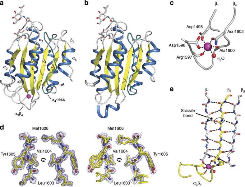
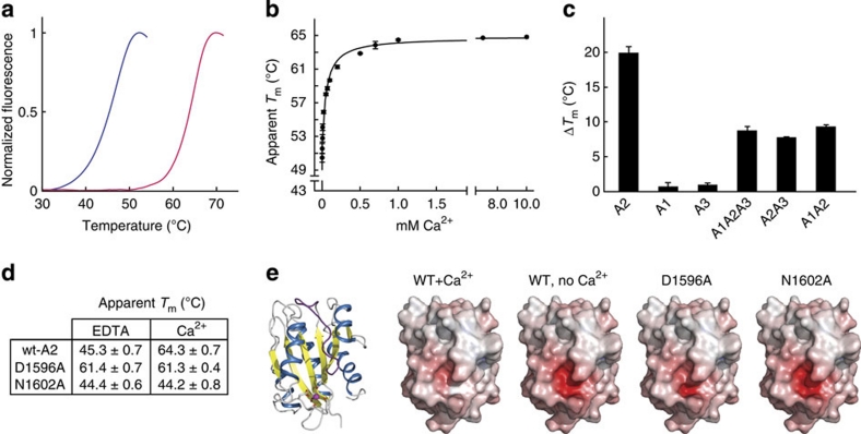
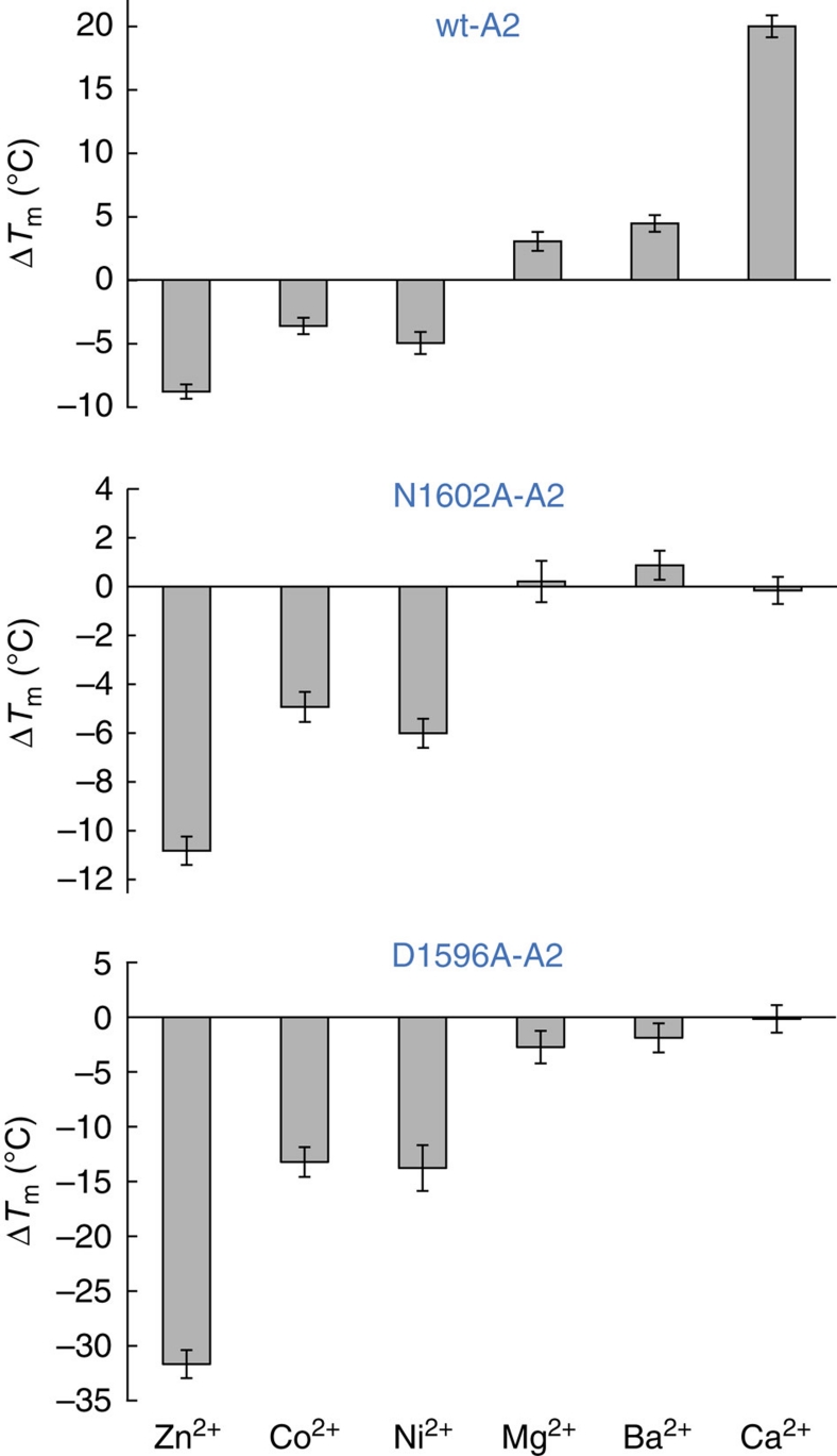
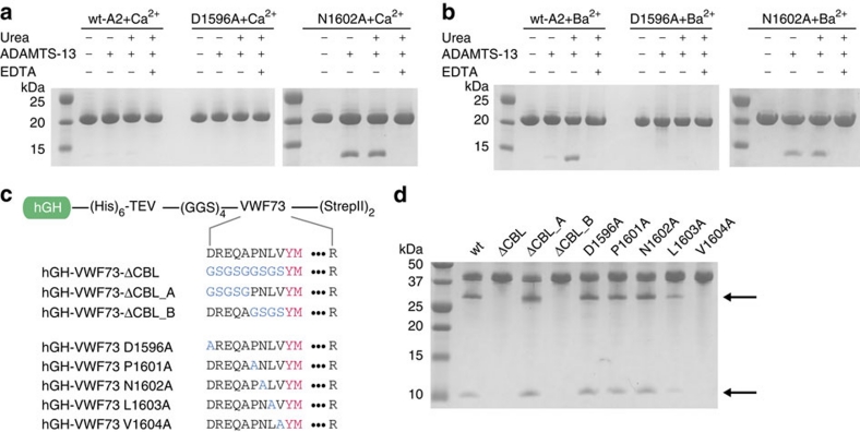
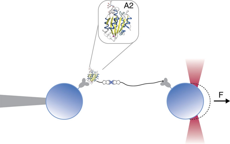
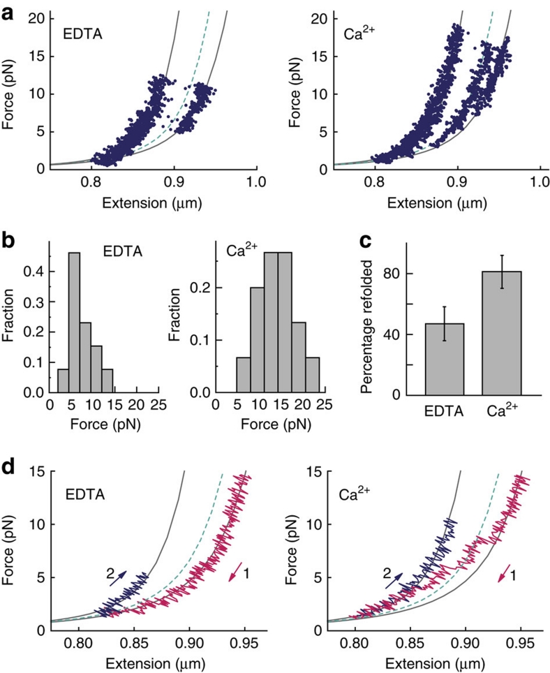
von Willebrand factor (VWF) multimers mediate primary adhesion and aggregation of platelets. VWF potency critically depends on multimer size, which is regulated by a feedback mechanism involving shear-induced unfolding of the VWF-A2 domain and cleavage by the metalloprotease ADAMTS-13. Here we report crystallographic and single-molecule optical tweezers data on VWF-A2 providing mechanistic insight into calcium-mediated stabilization of the native conformation that protects A2 from cleavage by ADAMTS-13. Unfolding of A2 requires higher forces when calcium is present and primarily proceeds through a mechanically stable intermediate with non-native calcium coordination. Calcium further accelerates refolding markedly, in particular, under applied load. We propose that calcium improves force sensing by allowing reversible force switching under physiologically relevant hydrodynamic conditions. Our data show for the first time the relevance of metal coordination for mechanical properties of a protein involved in mechanosensing.


Figures

Figure 1
Figure 1. The calcium-binding site of A2 affects scissile strand dynamics.
Crystal structures of calcium-bound wt-A2 (a) and, for comparison, calcium-free A2 (b; protein data bank (PDB) accession code 3GXB, ref. 12) are shown in cartoon representation. The central β-sheet (yellow) is encircled by α-helices (blue). Magenta, calcium ion; dark cyan, α4-less loop; sticks, N-acetyl-D-glucosamine (GlcNAG) on residues Asn1515 and Asn1574. Both structures are similar, except for the clear rearrangement of the α3β4-loop (a 310 helix in calcium-free A2) that forms most of the calcium-binding site. (c) Close-up of the calcium-binding site of A2. The calcium ion is coordinated by the α3β4-loop, Asp1498 and a water molecule (red sphere). Key side chains are shown in stick representation. (d) Close-ups of the scissile bond region in calcium-bound A2 (left) and calcium-free A2 (PDB accession code 3GXB, ref. ; right) show, respectively, a single, well-defined conformation and local disorder with multiple side and main-chain conformations. Electron density is contoured at 0.6 eÅ−3. (e) Backbone representation of the β4-scissile strand (yellow) and neighbouring strands illustrating how calcium interlocks β4 with β1 via coordination by Asp1498 and Asn1602.
Figure 2
Figure 2. Calcium stabilizes the native conformation of A2.
(a) Thermofluor stability assays were performed with monomeric A2 in buffer without (blue) or with (red) 1 mM CaCl2. Curves were normalized to maximum fluorescence signal. Data show representative curves of triplicate experiments. (b) Calcium binds A2 with high affinity and stabilizes A2 in a concentration-dependent manner. Apparent melting temperatures were extracted from the mid-point of the unfolding transitions. Error bars represent s.d. (c) Comparison of the difference in apparent melting temperature for individual VWF-A domains with native tandem constructs determined in the presence and absence of 1 mM CaCl2. A consistent shift of the unfolding temperature is only observed if A2 is part of the construct. Bar graphs represent mean Tm differences with respect to conditions containing EDTA (n=3). Error bars represent s.d. (d) Summary of unfolding temperatures (±s.d.) in the presence and absence of calcium. (e) Comparison of surface electrostatic potentials of wt-A2 and the D1596A and N1602A mutants. As reference, a ribbon model is shown in the same orientation. Electrostatic potentials were calculated on the solvent-accessible surface and contoured at −15 (red) to +15 (blue) kTe−1 (where k is the Boltzmann constant, T is temperature and e is elementary charge). The strongly negatively charged surface pocket forms the calcium-binding site. Thermal stability correlates with neutralization of electrostatic repulsion in the calcium-binding pocket.
Figure 3
Figure 3. Thermal stabilization of A2 is specific for calcium.
Thermofluor stability assays were performed with the monomeric wt-A2 or the calcium-binding-deficient mutants N1602A and D1596A in buffer with 1 mM CaCl2, 20 mM BaCl2, 10 mM MgCl2, 10 mM CoCl2, 10 mM NiCl2 or 10 mM ZnCl2. Bar graphs represent mean Tm differences with respect to conditions containing EDTA (n=3). The qualitative similarity of the data obtained for wt-A2 and both mutants with respect to the effect of Co2+, Ni2+ and Zn2+ indicates that the destabilizing effect observed for these metal ions does not result from competition with the calcium site. Error bars represent s.d.
Figure 4
Figure 4. VWF-A2 proteolysis by ADAMTS-13.
(a) The recombinant wt-A2 or the calcium-binding-deficient mutants D1596A and N1602A were incubated with 6 nM ADAMTS-13 and 5 mM CaCl2, with and without 1.5 M urea. No cleavage is observed under these conditions for wt-A2 and D1596A, whereas the destabilized N1602A mutant is cleaved efficiently. (b) ADAMTS-13 cleaves wt-A2 and N1602A, but not D1596A if BaCl2 replaces CaCl2. Because of divalent ion dependence of ADAMTS-13, no cleavage occurs in the presence of EDTA. (c) hGH–VWF73 fusion constructs. A (Gly–Gly–Ser)4 linker was engineered between hGH and VWF73 to ensure that substrate cleavage is not influenced by vicinity of the hGH fusion. Substituted fragments of the α3β4-loop are highlighted in blue; the Tyr1605–Met1606 cleavage site is shown in magenta. (d) ADAMTS-13 cleavage of hGH–VWF73 constructs. VWF73 proteins with D1596A, P1601A and N1602A mutations are cleaved at similar rates as the wt sequence. Substrate specificity resides in the P3 and P2 residues Leu1603 and Val1604. Arrows indicate N- (upper arrow) and C-terminal (lower arrow) cleavage products. TEV: cleavage site for tobacco etch virus protease; CBL: calcium-binding loop.
Figure 5
Figure 5. Schematic illustration of the optical tweezers experiment.
Single A2 domains (enlarged inset) are tethered between polystyrene beads that are held by a piezo-controlled micropipette and the optical trap. The tether includes a 2,500-bp DNA spacer to prevent unspecific bead–bead interactions.
Figure 6
Figure 6. Calcium modulates the folding and unfolding pathways of A2 under tensile force.
(a) Representative force-extension data showing six unfolding experiments, without (left) and with (right) calcium. Grey lines represent WLC fits to the native and unfolded states. The green dashed line is the WLC fit to a subset of the data that populates an intermediate state. (b) Force distribution histograms for unfolding events with and without calcium obtained at a loading rate of 5 pN s−1. Mean unfolding forces are 7±3 pN (n=13) and 14±4 pN (n=15), respectively. (c) Probability to refold to the native state at zero load significantly increases in the presence of calcium (P<0.03). T-bars represent s.d. from counting statistics. (d) Relaxation traces (magenta) of A2 molecules without (left) or with (right) calcium show that A2 is able to refold against load only when calcium is present. The dashed line is the WLC fit to the intermediate observed during unfolding. Stretching traces observed after successful refolding are shown in blue.

References

    1. Sadler J. E. Biochemistry and genetics of von Willebrand factor. Annu. Rev. Biochem. 67, 395–424 (1998). - PubMed
    1. Ruggeri Z. M. Von Willebrand factor. Curr. Opin. Hematol. 10, 142–149 (2003). - PubMed
    1. Wagner D. D. Cell biology of von Willebrand factor. Annu. Rev. Cell Biol. 6, 217–246 (1990). - PubMed
    1. Fowler W. E., Fretto L. J., Hamilton K. K., Erickson H. P. & McKee P. A. Substructure of human von Willebrand factor. J. Clin. Invest. 76, 1491–1500 (1985). - PMC - PubMed
    1. Furlan M., Robles R. & Lamie B. Partial purification and characterization of a protease from human plasma cleaving von Willebrand factor to fragments produced by in vivo proteolysis. Blood 87, 4223–4234 (1996). - PubMed

Publication types

Associated data