. 2011 Dec;19(12):1246-55.
doi: 10.1038/ejhg.2011.131. Epub 2011 Jul 13.

iPS cells to model CDKL5-related disorders

Affiliations

iPS cells to model CDKL5-related disorders

Mariangela Amenduni et al. Eur J Hum Genet. 2011 Dec.

Abstract

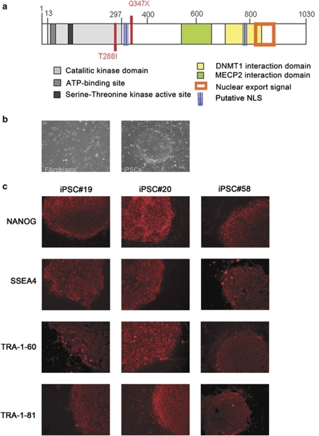
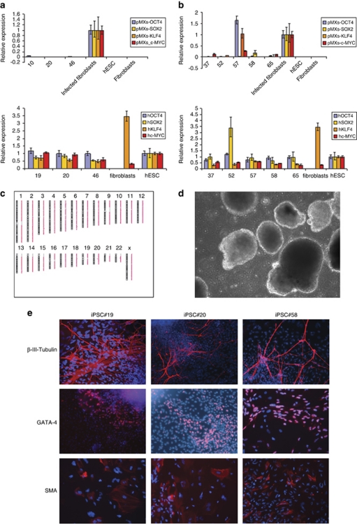
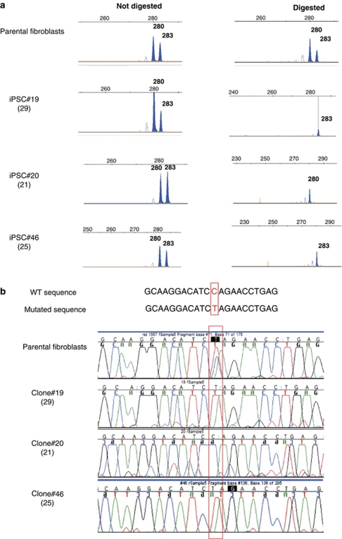
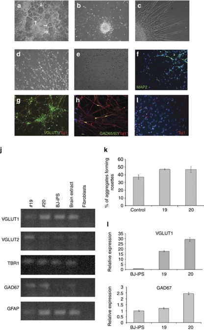
Rett syndrome (RTT) is a progressive neurologic disorder representing one of the most common causes of mental retardation in females. To date mutations in three genes have been associated with this condition. Classic RTT is caused by mutations in the MECP2 gene, whereas variants can be due to mutations in either MECP2 or FOXG1 or CDKL5. Mutations in CDKL5 have been identified both in females with the early onset seizure variant of RTT and in males with X-linked epileptic encephalopathy. CDKL5 is a kinase protein highly expressed in neurons, but its exact function inside the cell is unknown. To address this issue we established a human cellular model for CDKL5-related disease using the recently developed technology of induced pluripotent stem cells (iPSCs). iPSCs can be expanded indefinitely and differentiated in vitro into many different cell types, including neurons. These features make them the ideal tool to study disease mechanisms directly on the primarily affected neuronal cells. We derived iPSCs from fibroblasts of one female with p.Q347X and one male with p.T288I mutation, affected by early onset seizure variant and X-linked epileptic encephalopathy, respectively. We demonstrated that female CDKL5-mutated iPSCs maintain X-chromosome inactivation and clones express either the mutant CDKL5 allele or the wild-type allele that serve as an ideal experimental control. Array CGH indicates normal isogenic molecular karyotypes without detection of de novo CNVs in the CDKL5-mutated iPSCs. Furthermore, the iPS cells can be differentiated into neurons and are thus suitable to model disease pathogenesis in vitro.


Figures

Figure 1
Figure 1
Generation and characterization of iPSCs. (a) Schematic representation of CDKL5 protein structure with the known functional domains. The mutations identified in the patients reported in this study are shown in red. (b) Phase contrast image of human fibroblasts morphology before reprogramming (left) and typical morphology of one iPSC clone (right). Cells surrounding the clone are mitomycin-C-inactivated mouse embryo fibroblasts (feeders). (c) Representative images of immunostaining for pluripotency markers for two clones derived from patient 1 (iPSC#19 and iPSC#20) and one clone from patient 2 (iPSC#58). Images are at × 20 magnification.
Figure 2
Figure 2
iPSCs characterization. (a, b). Summary of real-time RT-PCR experiments demonstrating that clones derived from patient 1 (a) and from patient 2 (b) have inactivated the four transgenes (upper panel in a, b) and reactivated the corresponding endogenous genes (lower panel). A human ESC line and freshly infected fibroblasts were used as positive controls for the expression of endogenous genes and transgenes, respectively. Parental fibroblasts were also analyzed. Clone#57 from patient 2 maintained transgenes expression and it was thus excluded from further experiments. (c) Representative array CGH result from iPSC#19 from patient 1 showing a normal karyotype. (d) Phase contrast image of EBs after 5 days of suspension culture. (e) Immunostaining of EBs after 16 days of differentiation shows staining for markers specific of all three germ layers: β-III-tubulin (ectoderm), GATA-4 (endoderm) and smooth muscle actin (SMA; mesoderm). Representative images of two clones from patient 1 (iPSC#19 and iPSC#20) and one clone from patient 2 (iPSC#58) are shown. Images are at × 20 magnification.
Figure 3
Figure 3
X-inactivation analysis. (a) In parental fibroblasts two alleles can be identified. PCR on digested DNA still result in two peaks, indicating a balanced XCI pattern. Undigested DNA from the three iPS clones (#19, #20 and #46) present the same two peaks observed in parental fibroblasts. However, following digestion, each clone present only one peak, indicating a skewed pattern of XCI. Comparison of alleles length demonstrates that the three clones inactivate different X chromosomes. (b) Direct sequencing on cDNA from parental fibroblasts and iPSCs clones #19, #20 and #46 from patient 1. Wild-type and mutated sequences are shown at the top with the mutated nucleotide outlined in red. The chromatograms show that RNA isolated from parental fibroblasts presents both alleles whereas iPSCs clones express only one allele (mutated nucleotide outlined by a red rectangle). In particular, clones #19 and #46 express the mutated CDKL5 allele whereas clone #20 expresses the wt (wild-type) allele. Passage number at the moment of DNA and RNA extraction is indicated in parenthesis below clone number.
Figure 4
Figure 4
CDKL5-mutated iPSCs differentiate into neurons. iPSCs were induced to differentiate into neurons following a published protocol. (a) Neural rosettes (arrows) consisting of columnar cells arranged in a tubular structure were visible a few days after EBs seeding. (b) Shortly after neurospheres plating, numerous cell processes started to emerge from the spheres giving them a star-like appearance. (c, d) When cells were allowed to differentiate further, cell processes formed bundles of fibers (c) and cells could be seen migrating away from the spheres (d). (e, f) At this stage MAP2-positive neurons (green in f) could be identified by immunofluorescence. (g, h) After 10 weeks of differentiation the majority of β-III-tubulin-positive neurons (Tuj1, red) were also positive for VGLUT1 (green in g), but cells positive for the GABAergic marker GAD65/67 could be also identified (green in h). (i) In addition to neurons, many β-III-tubulin-negative non-neuronal cells were present in our cultures, as evidenced by DAPI-positive nuclei (blue). Images are at × 20 magnification. Blue staining=DAPI. (j) RT-PCR analysis on RNA isolated from 10-week-old neuronal cultures demonstrates that glutamatergic (VGLUT1, VGLUT2, TBR1) and GABAergic (GAD67) neurons, and glial cells (GFAP) are present in our cultures. RNA isolated from parental fibroblasts was used as negative control; a commercial RNA from human total brain was used as positive control. (k) The ability of our cells to differentiate toward a neuronal fate was estimated as the percentage of plated EBs that formed rosettes on day 15. Histograms represent the mean of two independent experiments. Error bars represent standard deviation. (l) Results of quantitative real-time RT-PCR on neuronal cultures show a high variability of expression of both VGLUT1 (upper panel; P<0.0001) and GAD67 (lower panel; P<0.0001 between BJ-iPS and #20, and between #19 and #20; P<0.05 between BJ-iPS and #19) in the three clones. Expression in neurons derived from the normal control clone was arbitrarily set as 1. Error bars represent standard deviation from three replicates of each sample.

References

    1. Chahrour M, Zoghbi HY. The story of Rett syndrome: from clinic to neurobiology. Neuron. 2007;56:422–437. - PubMed
    1. Hagberg BA, Skjeldal OH. Rett variants: a suggested model for inclusion criteria. Pediatr Neurol. 1994;11:5–11. - PubMed
    1. Rolando S. Rett syndrome: report of eight cases. Brain Dev. 1985;7:290–296. - PubMed
    1. Hanefeld F. The clinical pattern of the Rett syndrome. Brain Dev. 1985;7:320–325. - PubMed
    1. Zappella M. The preserved speech variant of the Rett complex: a report of 8 cases. Eur Child Adolesc Psychiatry. 1997;6:23–25. - PubMed

Publication types

Substances