. 2011 Jul;7(7):e1002091.
doi: 10.1371/journal.pgen.1002091. Epub 2011 Jul 7.

Genome-wide scan identifies TNIP1, PSORS1C1, and RHOB as novel risk loci for systemic sclerosis

Affiliations

Genome-wide scan identifies TNIP1, PSORS1C1, and RHOB as novel risk loci for systemic sclerosis

Yannick Allanore et al. PLoS Genet. 2011 Jul.

Abstract

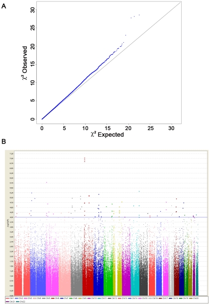
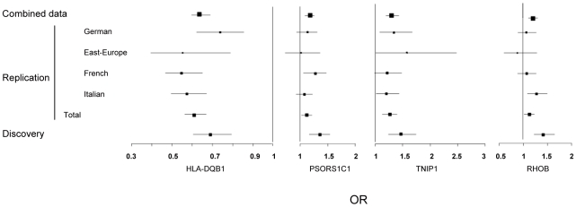
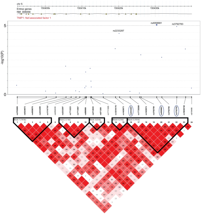
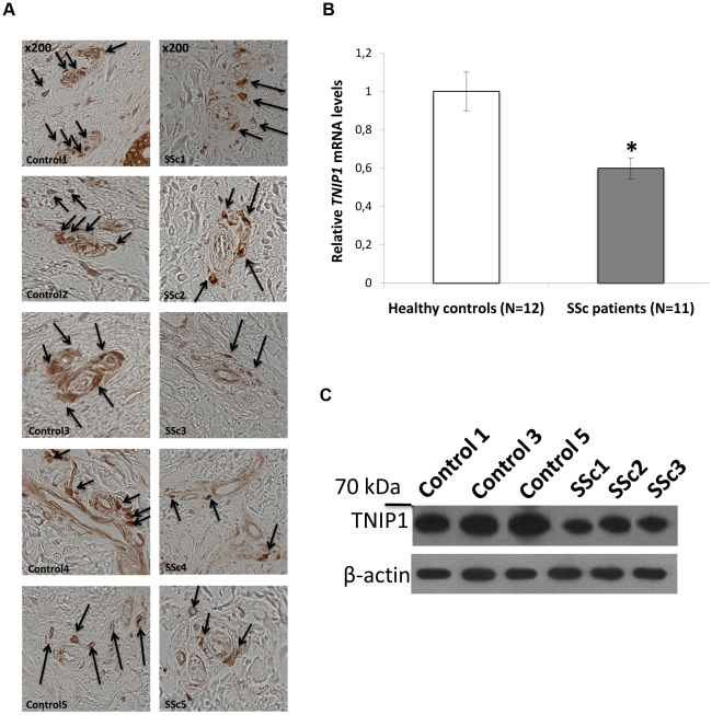
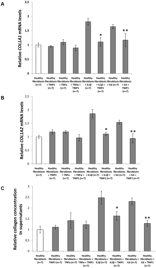
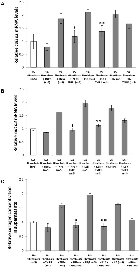
Systemic sclerosis (SSc) is an orphan, complex, inflammatory disease affecting the immune system and connective tissue. SSc stands out as a severely incapacitating and life-threatening inflammatory rheumatic disease, with a largely unknown pathogenesis. We have designed a two-stage genome-wide association study of SSc using case-control samples from France, Italy, Germany, and Northern Europe. The initial genome-wide scan was conducted in a French post quality-control sample of 564 cases and 1,776 controls, using almost 500 K SNPs. Two SNPs from the MHC region, together with the 6 loci outside MHC having at least one SNP with a P<10(-5) were selected for follow-up analysis. These markers were genotyped in a post-QC replication sample of 1,682 SSc cases and 3,926 controls. The three top SNPs are in strong linkage disequilibrium and located on 6p21, in the HLA-DQB1 gene: rs9275224, P = 9.18×10(-8), OR = 0.69, 95% CI [0.60-0.79]; rs6457617, P = 1.14×10(-7) and rs9275245, P = 1.39×10(-7). Within the MHC region, the next most associated SNP (rs3130573, P = 1.86×10(-5), OR = 1.36 [1.18-1.56]) is located in the PSORS1C1 gene. Outside the MHC region, our GWAS analysis revealed 7 top SNPs (P<10(-5)) that spanned 6 independent genomic regions. Follow-up of the 17 top SNPs in an independent sample of 1,682 SSc and 3,926 controls showed associations at PSORS1C1 (overall P = 5.70×10(-10), OR:1.25), TNIP1 (P = 4.68×10(-9), OR:1.31), and RHOB loci (P = 3.17×10(-6), OR:1.21). Because of its biological relevance, and previous reports of genetic association at this locus with connective tissue disorders, we investigated TNIP1 expression. A markedly reduced expression of the TNIP1 gene and also its protein product were observed both in lesional skin tissue and in cultured dermal fibroblasts from SSc patients. Furthermore, TNIP1 showed in vitro inhibitory effects on inflammatory cytokine-induced collagen production. The genetic signal of association with TNIP1 variants, together with tissular and cellular investigations, suggests that this pathway has a critical role in regulating autoimmunity and SSc pathogenesis.


Conflict of interest statement

The authors have declared that no competing interests exist.

Figures

Figure 1
Figure 1. Genome-wide association results from the discovery phase.
(A) Quantile-quantile plot for test statistics (logistic regression test) for 489,814 SNPs passing quality control. The plot shows a close match to the test statistics expected under the null distribution (λ = 1.03). (B) Manhattan plot representing the P values across the genome. The −log10 P of the logistic regression test (y axis) from 489,814 SNPs in 564 systemic sclerosis patients and 1,776 controls is plotted against its physical position (x-axis) on successive chromosomes. 90 SNPs with P<10−4 lie above the blue horizontal line and are listed in Table S1. Highly significant association was observed with SNPs within the MHC locus, including 1 SNPs that reached the conservative threshold for genome-wide significance (P<10−7).
Figure 2
Figure 2. Forest plots showing odds ratios and confidence intervals of the HLA-DQB1, PSORS1C1, TNIP1, and RHOB associations in the various populations studied in stage-1 and stage-2 data.
Figure 3
Figure 3. Association and linkage disequilibrium patterns at the TNIP1 gene.
(A) Association of SNPs in TNIP1: −log10 P of the logistic regression test for association (y axis) in the GWAS stage of SNPs is plotted against their physical position. The continuous line corresponds to P<10−5, the minimum P value of the top 7 SNPs identified in stage 1. The three SNPs that were followed and replicated in stage 2 are highlighted by a blue circle. Positions are given as NCBI build. (B) Linkage disequilibrim patterns at the TNIP1gene: pairwise LD (D′) are indicated by color gradients: D′≥0.80, red; 0.5≤D′<0.8, pink; 0.2≤D′<0.5, light pink; D′<0.2, white. The 3 SNPs are in strong LD (r2 = 0.57/0.72 between rs3792783 and rs2233287/rs495881). Intron and exon structure of the TNIP1 gene are taken from the UCSC Genome Browser.
Figure 4
Figure 4. Decreased TNIP1 expression in SSc patients.
(A) Expression of the TNIP1 protein was decreased ex vivo in SSc lesional skin tissue compared to controls (arrows indicate TNIP-1 positive cells). Shown are representative sections of the 5 patients and controls included in the analysis. (B) Consistent with these findings, a 1.7-fold decrease of TNIP1 mRNA levels was observed in dermal fibroblasts from SSc patients (* indicates a P-value = 0.001 vs. controls). These results were confirmed at the protein level (C).
Figure 5
Figure 5. TNIP1 abrogates the profibrotic effects of proinflammatory cytokines on collagen synthesis by healthy fibroblatsts.
(A,B) Healthy dermal fibroblasts treated with recombinant Il1β or Il6 and incubated 24 hours with TNIP1 displayed decreased mRNA levels for (A) COL1A1 (1.6 and 1.4-fold reduction, P = 0.01 and 0.03, respectively) and (B) COL1A2 (1.7 and 1.6-fold reduction, P = 0.02 and 0.03, respectively). No significant effect was observed in cells treated with recombinant TNFα. (C) Collagen content in cell culture supernatants treated with Il1β or Il6 was also reduced upon treatment with TNIP1 (1.5 and 1.8-fold reduction, P = 0.02 and 0.03, respectively). * indicates a P<0.05 versus healthy control fibroblasts treated with recombinant IL1β. ** indicates a P<0.05 versus healthy control fibroblasts treated with recombinant IL6.
Figure 6
Figure 6. TNIP1 abrogates the profibrotic effects of proinflammatory cytokines on collagen synthesis by SSc fibroblatsts.
(A,B) SSc dermal fibroblasts treated with recombinant TNFa or IL1β and incubated 24 hours with TNIP1 displayed decreased mRNA levels for (A) COL1A1 (1.6 and 1.5-fold reduction, P = 0.02 and 0.04, respectively) and (B) COL1A2 (1.7 and 1.8-fold reduction, P = 0.02 and 0.009, respectively). No significant effect was observed in cells treated with recombinant IL6. (C) Collagen content in cell culture supernatants treated with TNFa or IL1β was also reduced upon treatment with TNIP1 (1.8 and 2.3-fold reduction, P = 0.04 and 0.03, respectively). * indicates a P<0.05 vs. SSc fibroblasts treated with recombinant TNFa. ** indicates a P<0.05 vs. SSc fibroblasts treated with recombinant IL1 β.

References

    1. Valentini G, Black C. Systemic sclerosis. Best Pract Res Clin Rheumatol. 2002;16:807–16. - PubMed
    1. Thompson AE, Pope JE. Increased prevalence of scleroderma in southwestern Ontario: a cluster analysis. J Rheumatol. 29:1867–73. - PubMed
    1. Le Guern V, Mahr A, Mouthon L, Jeanneret D, Carzon M, et al. Prevalence of systemic sclerosis in a French multi-ethnic county. Rheumatology (Oxford) 2004;2004;43:1129–37. - PubMed
    1. Czirjak L, Kiss CG, Lovei C, Süto G, Varjú C, et al. Survey of Raynaud's phenomenon and systemic sclerosis based on a representative study of 10,000 south-Transdanubian Hungarian inhabitants. Clin Exp Rheumatol. 2005;23:801–8. - PubMed
    1. Arnett FC, Cho M, Chatterjee S, Aguilar MB, Reveille JD, et al. Familial occurrence frequencies and relative risks for systemic sclerosis (scleroderma) in three United States cohorts. Arthritis Rheum. 2001;44:1359–62. - PubMed

Publication types

MeSH terms