. 2011 Jul;7(7):e1002169.
doi: 10.1371/journal.pgen.1002169. Epub 2011 Jul 7.

NatF contributes to an evolutionary shift in protein N-terminal acetylation and is important for normal chromosome segregation

Affiliations

NatF contributes to an evolutionary shift in protein N-terminal acetylation and is important for normal chromosome segregation

Petra Van Damme et al. PLoS Genet. 2011 Jul.

Abstract

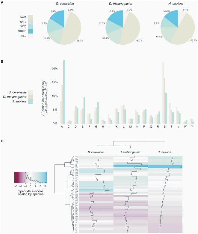
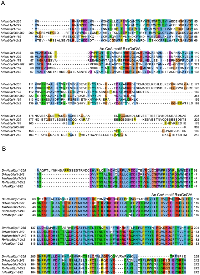
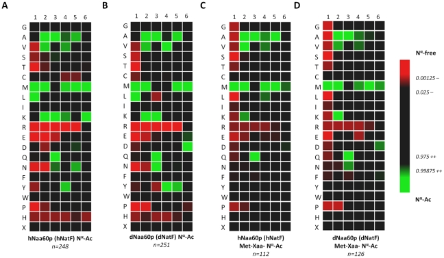
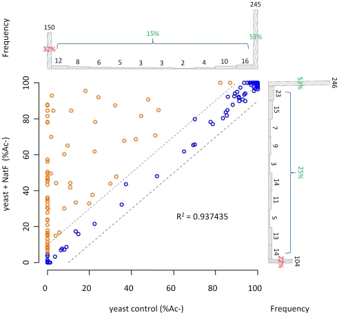
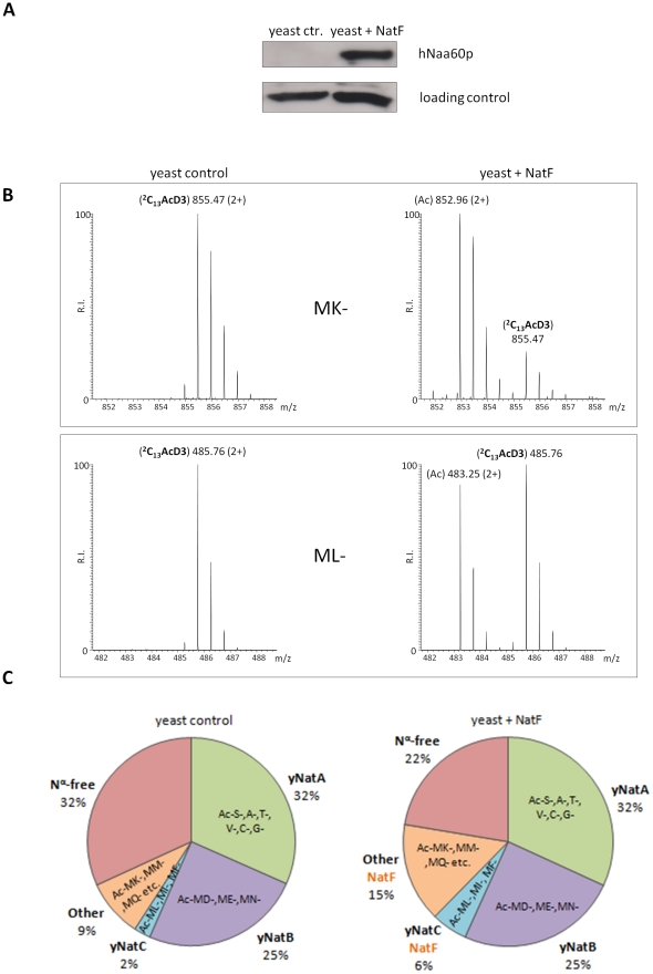
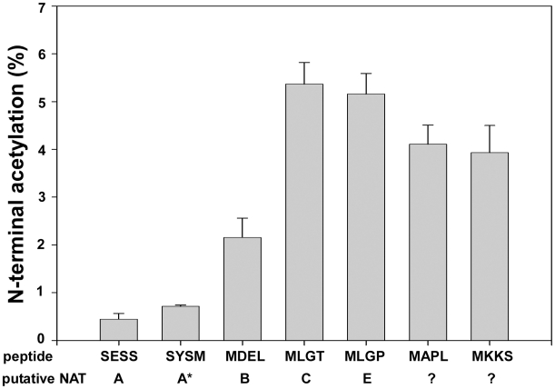
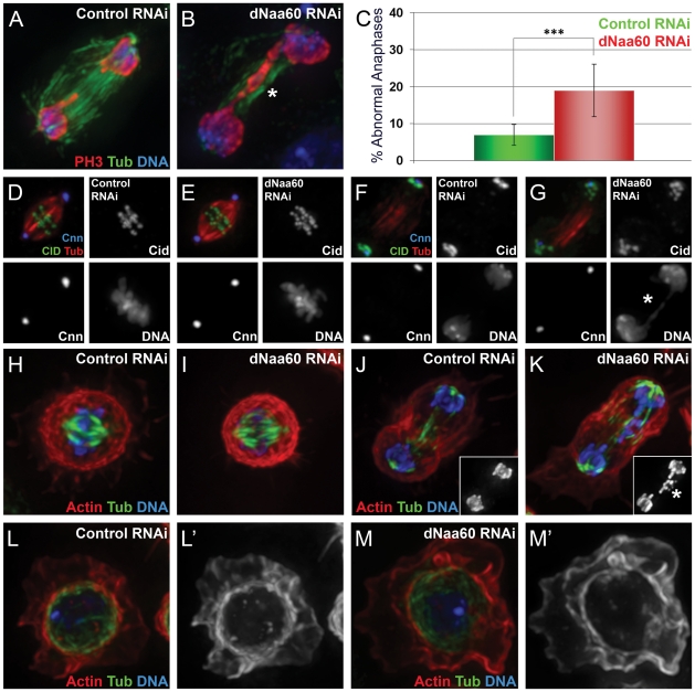
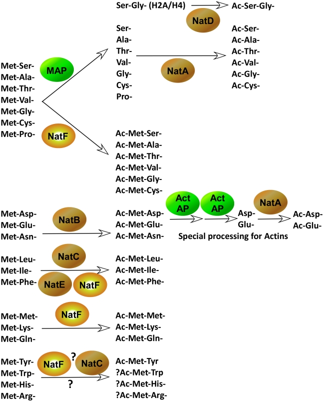
N-terminal acetylation (N-Ac) is a highly abundant eukaryotic protein modification. Proteomics revealed a significant increase in the occurrence of N-Ac from lower to higher eukaryotes, but evidence explaining the underlying molecular mechanism(s) is currently lacking. We first analysed protein N-termini and their acetylation degrees, suggesting that evolution of substrates is not a major cause for the evolutionary shift in N-Ac. Further, we investigated the presence of putative N-terminal acetyltransferases (NATs) in higher eukaryotes. The purified recombinant human and Drosophila homologues of a novel NAT candidate was subjected to in vitro peptide library acetylation assays. This provided evidence for its NAT activity targeting Met-Lys- and other Met-starting protein N-termini, and the enzyme was termed Naa60p and its activity NatF. Its in vivo activity was investigated by ectopically expressing human Naa60p in yeast followed by N-terminal COFRADIC analyses. hNaa60p acetylated distinct Met-starting yeast protein N-termini and increased general acetylation levels, thereby altering yeast in vivo acetylation patterns towards those of higher eukaryotes. Further, its activity in human cells was verified by overexpression and knockdown of hNAA60 followed by N-terminal COFRADIC. NatF's cellular impact was demonstrated in Drosophila cells where NAA60 knockdown induced chromosomal segregation defects. In summary, our study revealed a novel major protein modifier contributing to the evolution of N-Ac, redundancy among NATs, and an essential regulator of normal chromosome segregation. With the characterization of NatF, the co-translational N-Ac machinery appears complete since all the major substrate groups in eukaryotes are accounted for.


Conflict of interest statement

The authors have declared that no competing interests exist.

Figures

Figure 1
Figure 1. Overview of yeast, fruit fly, and human N-termini in NAT-classes and amino acid prevalence.
A. Comparative analyses of the distribution of all methionine-starting yeast (6561), fruit fly (3120) and human SwissProt entries (20238) (SwissProt version 2011-05) according to their Nat-type. For simplicity, methionine processing was assumed to occur for (M)A-, (M)S-, (M)T-, (M)V-, (M)C-, (M)G- and (M)P- starting N-termini, while the X-P- rule was used to assign unacetylated database entries . B. Bar charts of the amino acid occurrence at position 2 of yeast, fruit fly and human SwissProt protein entries. C. Heatmap of the ten highest and lowest ranking dipeptide z-scores across H. sapiens, D. melanogaster and S. cerevisiae. Z-scores are scaled by species, with the highest and lowest ranking z-score colored with the same intensity in blue and red respectively. The species (X-axis) and the dipeptides (Y-axis) were grouped by hierarchical clustering using the euclidian distance matrix of the z-scores.
Figure 2
Figure 2. Amino acid sequence alignments of hNaa60p and other NATs.
A. Amino acid sequence alignment of NAT15/hNaa60p and known human NATs. Only amino acid 200–362 of hNaa30p was included in the alignment. The consensus Acetyl Coenzyme A (AcCoA) binding motif RxxGxG/A, where x can be any amino acid, is indicated. T-Coffee (http://www.ebi.ac.uk/Tools/t-coffee/index.html) was used to make the alignment. Purple background indicates acidic residues, red indicates basic residues, orange indicates glycine, yellow indicates proline, blue indicates hydrophobic residues, green indicates polar residues, and turquoise indicates histidine and tyrosine. B. Amino acid sequence alignment of Naa60p from Drosophila melanogaster (Dm), Danio rerio (Dr), Mus musculus (Mm), Rattus norvegicus (Rn) and Homo sapiens (Hs). The consensus Acetyl Coenzyme A (AcCoA) binding motif RxxGxG/A, where x can be any amino acid, is indicated. Colour codes are used as in A.
Figure 3
Figure 3. Heatmap visualization reflecting the in vitro substrate specificity of hNaa60p and dNaa60p.
A. Heatmap of the 248 unique hNaa60p-specific oligopeptide substrates (353 substrate peptides in total). B. Heatmap of the 251 unique dNaa60p-specific oligopeptide substrates (345 substrate peptides in total). C. Heatmap of the subset of 112 unique hNaa60p-specific methionine-starting oligopeptide substrates. D. Heatmap of the subset of 126 unique dNaa60p-specific methionine-starting oligopeptide substrates. Data was normalized against the natural positional amino acid composition of SwissProt (version 57.8) [iterative rounds (n = 100) of randomly selected sequences (n = 100) were taken as to correct for the statistical variations (SD = standard deviation) intrinsically present at each position in the experimental datasets ranging from amino acid 1 to 6]. The significance threshold was set at 0.01. Red color shades are negatively correlated with the occurrence in Naa60p peptide-substrates as compared to random sequences in SwissProt, while green shades are positively correlated.
Figure 4
Figure 4. NatF N-terminally acetylates yeast substrates in vivo.
Scatterplot displaying the correlation of the degrees of Nα-acetylation when comparing a control (X-axis) and a human NatF (hNaa60p)-expressing (Y-axis) yeast N-terminome dataset. The correlation was calculated with the R statistical package to be R2 = 0.937. The N-termini displaying a significant variation in the degree of Nα-acetylation (see Materials and Methods) are highlighted in orange. The frequency histograms of the number of matching data points are also shown.
Figure 5
Figure 5. NatF expression shifts the overall status of the yeast N-terminal acetylome.
A. A yeast strain expressing hNaa60p/NatF ‘Yeast+NatF’ was generated (see Materials and Methods), and processed by SDS-PAGE and Western blotting along with the control strain ‘Yeast ctr.’ containing an empty control plasmid. Anti-hNaa60p verified expression of hNaa60p in the Yeast+NatF strain (an unspecific band served as loading control). B. MS-spectra from the MK- starting N-terminal peptide (doubly charged precursor) of the Smr domain-containing protein YPL199C (1MKGTGGVVVGTQNPVR16) reveals two distinguishable isotopic envelopes in the hNaa60p expressing yeast strain [i.e. the acetylated (Ac) and 2C13 and trideutero-acetylated forms (2C13AcD3), right upper panel] indicative for the fact that this N-terminus is 82% in vivo Nα-acetylated while being Nα-free in the control sample (left upper panel). The lower panels show MS-spectra of the ML-starting N-terminal peptide (doubly charged precursor) of the uncharacterized protein YGR130C (1MLFNINR7) in the control sample (0% Nα-acetylated, left lower panel) or hNAA60 sample (48% acetylated, right lower panel). C. Nat-category specific distribution of experimentally identified yeast N-termini in the yeast control or hNaa60p-expressing yeast strain. Only those N-termini of which the N-Ac status could univocally be assigned (n = 464) were considered.
Figure 6
Figure 6. Knockdown and overexpression of hNaa60p affects N-terminal acetylation in HeLa cells.
A. HeLa cells cultivated in 13C6 15N4 L-arginine were transfected with control vector and cells cultivated in 12C6 L-arginine were transfected with phNAA60-V5. After 48 hours of transfection the cells were harvested, lysed and subjected to COFRADIC and MS and MS/MS- analysis. MS spectra of the peptide 1MKGKEEKEGGAR12, originating from the STIP1 homology and U-box containing protein1 is shown. The protein is more acetylated when hNaa60p is overexpressed (32% Nα-acetylated) as compared to the control (18% Nα-acetylated). Aliquots were processed by SDS-PAGE and Western blotting using anti-V5 and anti-β-tubulin antibodies. B. Control cells cultivated in 12C6 L-arginine were transfected with 50 nM siNon-targeting control, and cells cultivated in 13C6 15N4 L-arginine were transfected with 50 nM sihNAA60 pool. After 84 hours of transfection the cells were harvested and subjected to COFRADIC and MS analysis. MS spectra of the peptide 1MVPGSEGPAR10, originating from the protein Mediator of RNA polymerase II transcription subunit 25 is shown. The peptide was partially acetylated in both control (26% Nα-acetylated) and knockdown setup (17% Nα-acetylated), however the peptide was less Nα-acetylated when the levels of hNaa60p was reduced. Aliquots were processed for SDS-PAGE and Western blotting using anti-hNaa60p and anti-actin antibodies.
Figure 7
Figure 7. NAT-activity of recombinant hNaa60p towards synthetic N-terminal oligopeptides.
MBP-hNaa60p was incubated with the indicated oligopeptide substrates (200 µM) and acetyl-Coenzyme A (300 µM) in acetylation buffer for 35 minutes at 37°C. Peptide acetylation was determined by RP-HPLC peptide separation. The NATs responsible for acetylating the different peptides are shown. Question marks indicate that no NAT has yet been identified to acetylate these peptides. *SYSM represents the ACTH N-terminus which is an artificial in vitro substrate of NatA. The four first amino acids in the oligopeptides are indicated, for further details see Materials and Methods.
Figure 8
Figure 8. dNaa60p is required for chromosome segregation during anaphase.
Control dsRNA treated cells (A,D,F,H,J and L). dNAA60 dsRNA treated cells (B,E,G,I,K and M). dNAA60-depleted cells exhibited chromosome segregation defects during anaphase (A–C). These defects included lagging chromosomes (K, highlighted by asterisk) and chromosome bridges (B and G, highlighted by asterisk). Quantification of chromosome segregation defects in dNAA60-depleted cells (n = 278) and control cells (n = 179) (***p<0,001 Student's test) (C). Histone 3 phosphorylated on Serine 10 (red), α-tubulin (green) and DNA (blue). dNAA60-depleted cells showed no significant defects in the localization of both Cnn and Cid proteins (D–G). Both control and dNAA60-depleted cells exhibited bypolar spindles with correct alignment of chromosomes at the metaphase plate (D,E). Anaphase cells with chromosome segregation defects in dNAA60-depleted cells showed no obvious defects in Cnn and Cid localization (F,G). α-tubulin (red), Cid (green) and Cnn (blue). Control and dNAA60-depleted cells exhibit proper chromosome alignment during metaphase with no detectable defects in the actin and microtubule cytoskeleton (H,I). dNAA60-depleted cells undergoing anaphase with chromosome segregation defects also showed a normal actin and microtubule cytoskeleton (J,K; details show histone 3 phosphorylated on serine 10 staining). dNAA60-depleted cells in interphase show no detectable defects regarding the actin and microtuble cytoskeleton (L–M). Actin (red), α-Tubulin (green) and Histone 3 phosphorylated on Serine 10 (blue).
Figure 9
Figure 9. The major pathways of protein N-terminal processing in higher eukaryotes.
N-termini of which the iMet is followed by one of the small amino acids, Ser-, Ala-, Thr-, Val-, Gly-, Cys-, and Pro- often undergo iMet cleavage performed by a methionine aminopeptidase (MAP). These N-termini, with the exception of Pro-, are often further acetylated by NatA, or in the case of Histone H2A and H4, by NatD (Hole K. et al., unpublished). However, this group of N-termini may also be acetylated by NatF. Met-Asp-, Met-Glu- and Met-Asn- are acetylated by NatB. Actins are further processed in one or more steps by unidentified Actin aminopeptidases (Act AP). The acidic actin N-termini are then acetylated by at NAT, presumably NatA . Hydrophobic Met-Leu-, Met-Ile- and Met-Phe- are acetylated by NatC, but also by NatF as well as by NatE in vitro, suggesting potential redundancy between these NATs. Met-Met-, Met-Lys- and Met-Gln- are acetylated by NatF and potentially other NATs. Information about Met-His-, Met-Arg-, Met-Trp- and Met-Tyr- is limited, but it is likely that some of these N-termini are acetylated as well, by NatF and perhaps NatC.

References

    1. Behnia R, Panic B, Whyte JR, Munro S. Targeting of the Arf-like GTPase Arl3p to the Golgi requires N-terminal acetylation and the membrane protein Sys1p. Nat Cell Biol. 2004;6:405–413. - PubMed
    1. Caesar R, Blomberg A. The stress-induced Tfs1p requires NatB-mediated acetylation to inhibit carboxypeptidase Y and to regulate the protein kinase A pathway. J Biol Chem. 2004;279:38532–38543. - PubMed
    1. Coulton AT, East DA, Galinska-Rakoczy A, Lehman W, Mulvihill DP. The recruitment of acetylated and unacetylated tropomyosin to distinct actin polymers permits the discrete regulation of specific myosins in fission yeast. J Cell Sci. 2010;123:3235–3243. - PMC - PubMed
    1. Setty SR, Strochlic TI, Tong AH, Boone C, Burd CG. Golgi targeting of ARF-like GTPase Arl3p requires its Nalpha-acetylation and the integral membrane protein Sys1p. Nat Cell Biol. 2004;6:414–419. - PubMed
    1. Hwang CS, Shemorry A, Varshavsky A. N-terminal acetylation of cellular proteins creates specific degradation signals. Science. 2010;327:973–977. - PMC - PubMed

Publication types

MeSH terms