. 2011 Nov;112(11):3364-72.
doi: 10.1002/jcb.23267.

Zoledronic acid inhibits macrophage SOCS3 expression and enhances cytokine production

Affiliations

Zoledronic acid inhibits macrophage SOCS3 expression and enhances cytokine production

Erica L Scheller et al. J Cell Biochem. 2011 Nov.

Abstract

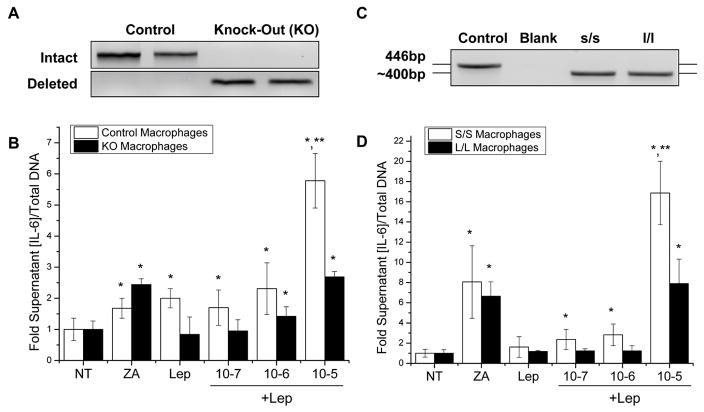
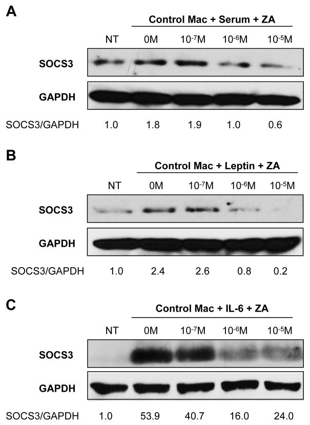
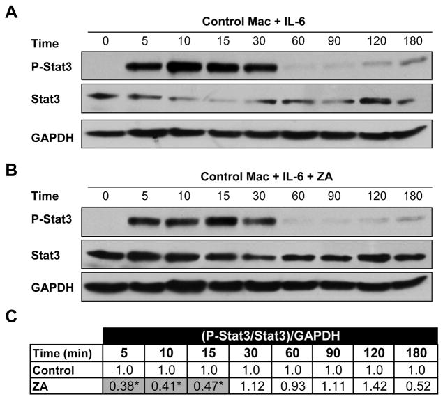
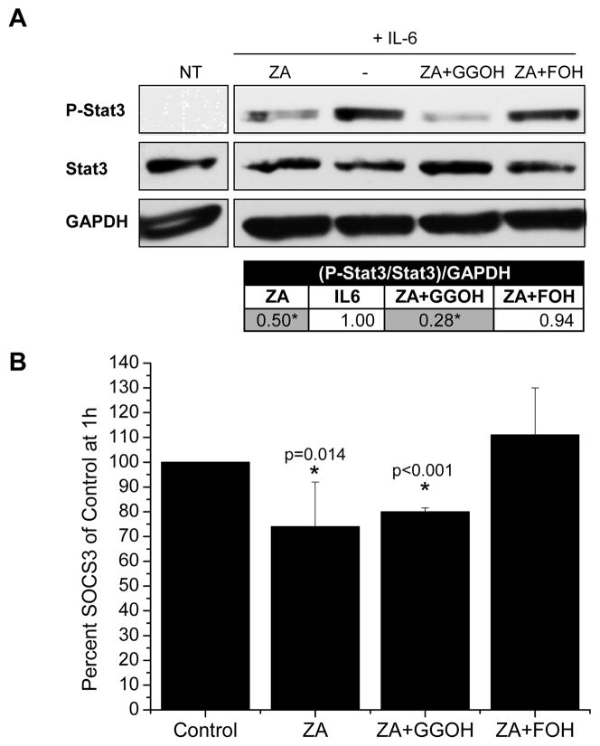
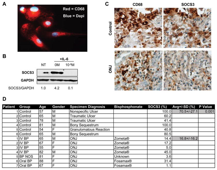
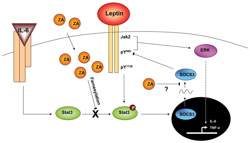
Suppressor of cytokine signaling-3 (SOCS3) has multiple functions including inhibition of Janus kinase (Jak) activity, regulation of protein degradation, and suppression of cytokine signaling. SOCS3 modulates macrophage response to cytokines such as IL-6 and leptin that are systemically induced in obesity. Obesity is a suspected risk factor for SOCS3-related pathology such as rheumatoid arthritis and Crohn's disease as well as zoledronic acid (ZA)-induced osteonecrosis of the jaw (ONJ). Thus, understanding the ability of bisphosphonates to modulate SOCS3 is necessary to qualify their contribution to these disorders. ONJ occurs in up to 10% of patients using intravenous bisphosphonates and has an unknown pathogenesis that may be linked to decreased bone turnover, altered vascularity, bacterial invasion, and compromised wound healing. Given the increased risk of ONJ with obesity and importance of macrophages in wound healing, we hypothesized that amino-bisphosphonates could contribute to the pathogenesis of ONJ by regulating macrophage responses to cytokines such as leptin and IL-6. We report that ZA is a novel inhibitor of SOCS3 in primary macrophages and human ONJ biopsy specimens. Inhibition of SOCS3 by ZA resulted in significant increases in IL-6 production. SOCS3 transcription is regulated by nuclear accumulation of phosphorylated-Stat3 (P-Stat3). We found that ZA decreased phosphorylation of Stat3 in a mevalonate-pathway dependent manner. However, restoration of P-Stat3 was not sufficient to correct SOCS3 inhibition. We propose that disruption of macrophage SOCS3 expression by amino-bisphosphonates such as ZA may be a novel contributor to inflammatory phenotypes in obesity and the pathogenesis of ONJ.


Figures

Figure 1
Figure 1. ZA and leptin induce synergistic downstream cytokine production mediated by ObRb tyrosine 985
(A) Genomic DNA PCR confirming complete cre-lox recombination of the BMMs from the long-form leptin-receptor (ObRb) knock-out (KO) animals and intact ObRb in the controls. (B) IL-6 ELISA of control and KO BMMs after 16–18 hour pretreatment with ZA followed by induction with 100nM leptin for 24 hours. (C) Confirmatory genotyping of s/s (point mutation ObRb Tyr1138) and l/l (point mutation ObRb Tyr985) mice. (D) IL-6 ELISA of s/s and l/l BMMs as above. *: significant over NT control, **: significant over ZA only control. (N=6, all experiments repeated at least twice)
Figure 2
Figure 2. ZA inhibits SOCS3 protein accumulation in primary bone marrow macrophages
Western blot densitometry data was normalized to GAPDH control. (A,B,C) ZA pretreatment for 16–18 hours from 10−7 to 10−5M dose-dependently inhibited induction of SOCS3 in BMMs by 20% serum, 100nM leptin, or 25ng/mL IL-6 respectively after three hours. Representative blots shown, all experiments repeated at least twice.
Figure 3
Figure 3. ZA inhibits Stat3 phosphorylation
(A) Control western blot of P-Stat3, Stat3, and GAPDH after BMM stimulation with 25ng/mL IL-6 for 0 to 180 minutes. (B) Paired western blot after treatment with 10−5M ZA. (C) Statistical analysis of the ratio of phosphorylation of Stat3 after ZA treatment to control at each time point (N=3). Control densitometry ratio [(P-Stat3/Stat3)/GAPDH] was set to 1.0 and corresponding ZA treated ratio given a proportional value before statistical comparison of independent blots.
Figure 4
Figure 4. Farnesyl intermediates rescue ZA inhibition of P-Stat3 and SOCS3 mRNA
(A) Western blot of BMMs pre-treated with 10−5M ZA and 10 μM GGOH or FOH overnight, ‘−’ indicates lack of ZA treatment. Pre-treated cells were stimulated with 25ng/mL IL-6 for 10–15 minutes. Tabulated results report the ratio of phosphorylation of Stat3 after ZA treatment to control at each time point as in figure 3 (N=3). (B) Quantitative PCR analysis of SOCS3 mRNA after one hour, treatments as in (B). Results are reported as percent of control SOCS3 mRNA (N=3–5). *: significantly less than control.
Figure 5
Figure 5. Rescue of P-Stat3 and SOCS3 mRNA with FOH does not correct SOCS3 protein inhibition
Representative western blot of SOCS3 after ZA/GGOH/FOH overnight pre-treatment where indicated and three hour 25ng/mL IL-6 stimulation of BMMs. GGOH and FOH concentrations are reported as μM and combined with 10−5M ZA. Experiment was repeated three times with similar results.
Figure 6
Figure 6. Bisphosphonates decrease SOCS3 in human CD68+ macrophages and human ONJ biopsies
(A) Cellular immunofluorescence of primary human macrophages. Red = CD68, Blue = Dapi. 40x magnification. (B) Representative western blot of primary macrophage SOCS3 after treatment with 50ng/mL IL-6 alone or IL-6 + 10−5M ZA. Experiment was performed in duplicate with similar results. (C) Representative serially stained sections of control ulcerated tissue and an ONJ lesion. 40x magnification. (D) Patient information, condition, treatment, and SOCS3 percent staining density.
Figure 7
Figure 7. Theoretical model of ZA regulation of SOCS3
ZA taken up by the cell blocks phosphorylation of Stat3 in a farnesylation-dependent manner. ZA, via a secondary mechanism that may involve inhibition of protein translation, blocks protein accumulation of SOCS3. Functionally, this results in increased cytokine output of leptin-induced IL-6 in an ObRb Tyr985, Erk signaling dependent manner.

References

    1. Banks AS, Davis SM, Bates SH, Myers MG., Jr Activation of downstream signals by the long form of the leptin receptor. J Biol Chem. 2000;275:14563–72. - PubMed
    1. Bates SH, Stearns WH, Dundon TA, Schubert M, Tso AW, Wang Y, Banks AS, Lavery HJ, Haq AK, Maratos-Flier E, Neel BG, Schwartz MW, Myers MG., Jr STAT3 signalling is required for leptin regulation of energy balance but not reproduction. Nature. 2003;421:856–9. - PubMed
    1. Bjornholm M, Munzberg H, Leshan RL, Villanueva EC, Bates SH, Louis GW, Jones JC, Ishida-Takahashi R, Bjorbaek C, Myers MG., Jr Mice lacking inhibitory leptin receptor signals are lean with normal endocrine function. J Clin Invest. 2007;117:1354–60. - PMC - PubMed
    1. Cartsos VM, Zhu S, Zavras AI. Bisphosphonate use and the risk of adverse jaw outcomes: a medical claims study of 714,217 people. J Am Dent Assoc. 2008;139:23–30. - PubMed
    1. Chang MK, Raggatt LJ, Alexander KA, Kuliwaba JS, Fazzalari NL, Schroder K, Maylin ER, Ripoll VM, Hume DA, Pettit AR. Osteal tissue macrophages are intercalated throughout human and mouse bone lining tissues and regulate osteoblast function in vitro and in vivo. J Immunol. 2008;181:1232–44. - PubMed

Publication types

MeSH terms