Review
. 2011 Aug;21(4):509-16.
doi: 10.1016/j.sbi.2011.06.001.

Femtosecond nanocrystallography using X-ray lasers for membrane protein structure determination

Affiliations
Review

Femtosecond nanocrystallography using X-ray lasers for membrane protein structure determination

Petra Fromme et al. Curr Opin Struct Biol. 2011 Aug.

Abstract

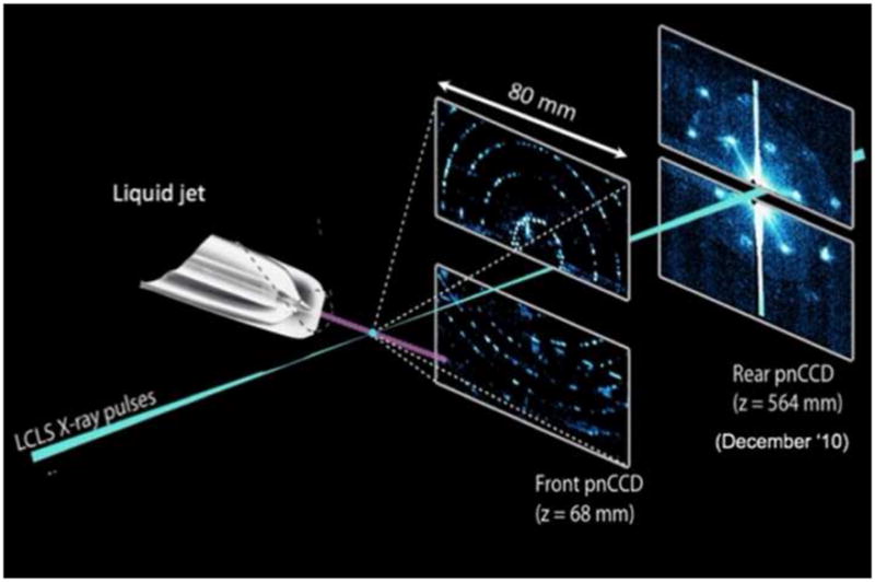
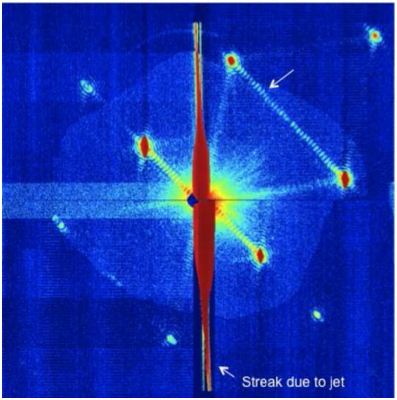
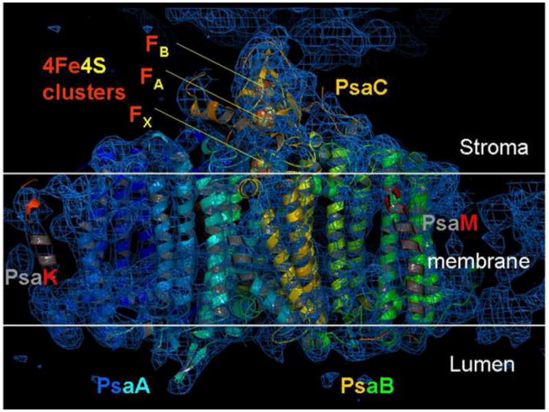
The invention of free electron X-ray lasers has opened a new era for membrane protein structure determination with the recent first proof-of-principle of the new concept of femtosecond nanocrystallography. Structure determination is based on thousands of diffraction snapshots that are collected on a fully hydrated stream of nanocrystals. This review provides a summary of the method and describes how femtosecond X-ray crystallography overcomes the radiation-damage problem in X-ray crystallography, avoids the need for growth and freezing of large single crystals while offering a new method for direct digital phase determination by making use of the fully coherent nature of the X-ray beam. We briefly review the possibilities for time-resolved crystallography, and the potential for making 'molecular movies' of membrane proteins at work.


Figures

Figure 1
Figure 1
Principle of the experimental setup used for femtosecond crystallography.. The stream of crystals is provided by the injector with the gas focusing nozzle. The femtosecond X-ray beam intersect with the continous crystal stream; diffraction patterns are recorded with the frequency of the Free Electron Laser at LCLS (60 or 120 Hz) using either a one or two CCD detector setup. In the two detector setup, which was used in the CAMP camber at the AMO beamline at LCLS for the first fs crystallography experiments reported in [•• 24], the high order reflections are detected on the front detector. The back detector, which records the low order reflections, allows the resolution of the shape transforms of the reflections. The figure has been modified after [•• 24].
Figure 2
Figure 2
Example of a diffraction pattern from photosystem I which shows the shape transforms. Note the fringes between the Bragg peaks which indicate the number of unit cells in that direction. Thus the evaluation of the shape transforms can be used to determine the size of the crystals [•• 24], and for phase determination [•• 38]. This Figure has been modified after [•• 24].
Figure 3
Figure 3
Electron density map of Photosystem I at 8 Åresolution, derived by fs crystallography. The phases were obtained using molecular replacement as described in [•• 24]. The section of the electron density in this picture shows transmembrane helices of the large PSI subunits PsaA, PsaB as well as helices of PsaK and PsaF. This Figure has been modified after [•• 25].

Similar articles

  • Femtosecond X-ray protein nanocrystallography.
    Chapman HN, Fromme P, Barty A, White TA, Kirian RA, Aquila A, Hunter MS, Schulz J, DePonte DP, Weierstall U, Doak RB, Maia FR, Martin AV, Schlichting I, Lomb L, Coppola N, Shoeman RL, Epp SW, Hartmann R, Rolles D, Rudenko A, Foucar L, Kimmel N, Weidenspointner G, Holl P, Liang M, Barthelmess M, Caleman C, Boutet S, Bogan MJ, Krzywinski J, Bostedt C, Bajt S, Gumprecht L, Rudek B, Erk B, Schmidt C, Hömke A, Reich C, Pietschner D, Strüder L, Hauser G, Gorke H, Ullrich J, Herrmann S, Schaller G, Schopper F, Soltau H, Kühnel KU, Messerschmidt M, Bozek JD, Hau-Riege SP, Frank M, Hampton CY, Sierra RG, Starodub D, Williams GJ, Hajdu J, Timneanu N, Seibert MM, Andreasson J, Rocker A, Jönsson O, Svenda M, Stern S, Nass K, Andritschke R, Schröter CD, Krasniqi F, Bott M, Schmidt KE, Wang X, Grotjohann I, Holton JM, Barends TR, Neutze R, Marchesini S, Fromme R, Schorb S, Rupp D, Adolph M, Gorkhover T, Andersson I, Hirsemann H, Potdevin G, Graafsma H, Nilsson B, Spence JC. Chapman HN, et al. Nature. 2011 Feb 3;470(7332):73-7. doi: 10.1038/nature09750. Nature. 2011. PMID: 21293373 Free PMC article.
  • Toward structure determination using membrane-protein nanocrystals and microcrystals.
    Hunter MS, Fromme P. Hunter MS, et al. Methods. 2011 Dec;55(4):387-404. doi: 10.1016/j.ymeth.2011.12.006. Epub 2011 Dec 22. Methods. 2011. PMID: 22197730 Free PMC article. Review.
  • Time-resolved protein nanocrystallography using an X-ray free-electron laser.
    Aquila A, Hunter MS, Doak RB, Kirian RA, Fromme P, White TA, Andreasson J, Arnlund D, Bajt S, Barends TR, Barthelmess M, Bogan MJ, Bostedt C, Bottin H, Bozek JD, Caleman C, Coppola N, Davidsson J, DePonte DP, Elser V, Epp SW, Erk B, Fleckenstein H, Foucar L, Frank M, Fromme R, Graafsma H, Grotjohann I, Gumprecht L, Hajdu J, Hampton CY, Hartmann A, Hartmann R, Hau-Riege S, Hauser G, Hirsemann H, Holl P, Holton JM, Hömke A, Johansson L, Kimmel N, Kassemeyer S, Krasniqi F, Kühnel KU, Liang M, Lomb L, Malmerberg E, Marchesini S, Martin AV, Maia FR, Messerschmidt M, Nass K, Reich C, Neutze R, Rolles D, Rudek B, Rudenko A, Schlichting I, Schmidt C, Schmidt KE, Schulz J, Seibert MM, Shoeman RL, Sierra R, Soltau H, Starodub D, Stellato F, Stern S, Strüder L, Timneanu N, Ullrich J, Wang X, Williams GJ, Weidenspointner G, Weierstall U, Wunderer C, Barty A, Spence JC, Chapman HN. Aquila A, et al. Opt Express. 2012 Jan 30;20(3):2706-16. doi: 10.1364/OE.20.002706. Opt Express. 2012. PMID: 22330507 Free PMC article.
  • Femtosecond crystallography of membrane proteins in the lipidic cubic phase.
    Liu W, Wacker D, Wang C, Abola E, Cherezov V. Liu W, et al. Philos Trans R Soc Lond B Biol Sci. 2014 Jul 17;369(1647):20130314. doi: 10.1098/rstb.2013.0314. Philos Trans R Soc Lond B Biol Sci. 2014. PMID: 24914147 Free PMC article. Review.
  • Diffraction by nanocrystals.
    Chen JP, Millane RP. Chen JP, et al. J Opt Soc Am A Opt Image Sci Vis. 2013 Dec 1;30(12):2627-34. doi: 10.1364/JOSAA.30.002627. J Opt Soc Am A Opt Image Sci Vis. 2013. PMID: 24323025

Cited by

References

    1. Cusack S, Belrhali H, Bram A, Burghammer M, Perrakis A, Riekel C. Small is beautiful: protein micro-crystallography. Nat Struct Biol. 1998;5 (Suppl):634–637. - PubMed
    1. Cherezov V, Caffrey M. Membrane protein crystallization in lipidic mesophases. A mechanism study using X-ray microdiffraction. Faraday Discuss. 2007;136:195–212. discussion 213–129. - PubMed
    1. Cherezov V, Rosenbaum DM, Hanson MA, Rasmussen SG, Thian FS, Kobilka TS, Choi HJ, Kuhn P, Weis WI, Kobilka BK, et al. High-resolution crystal structure of an engineered human beta2-adrenergic G protein-coupled receptor. Science. 2007;318:1258–1265. - PMC - PubMed
    1. Rasmussen SG, Choi HJ, Rosenbaum DM, Kobilka TS, Thian FS, Edwards PC, Burghammer M, Ratnala VR, Sanishvili R, Fischetti RF, et al. Crystal structure of the human beta2 adrenergic G-protein-coupled receptor. Nature. 2007;450:383–387. - PubMed
    1. Rosenbaum DM, Cherezov V, Hanson MA, Rasmussen SG, Thian FS, Kobilka TS, Choi HJ, Yao XJ, Weis WI, Stevens RC, et al. GPCR engineering yields high-resolution structural insights into beta2-adrenergic receptor function. Science. 2007;318:1266–1273. - PubMed

Publication types

Substances

LinkOut - more resources