Review
. 2011 Dec;42(8):762-72.
doi: 10.1016/j.micron.2011.06.005. Epub 2011 Jun 25.

The use of trehalose in the preparation of specimens for molecular electron microscopy

Affiliations
Review

The use of trehalose in the preparation of specimens for molecular electron microscopy

Po-Lin Chiu et al. Micron. 2011 Dec.

Abstract

Biological specimens have to be prepared for imaging in the electron microscope in a way that preserves their native structure. Two-dimensional (2D) protein crystals to be analyzed by electron crystallography are best preserved by sugar embedding. One of the sugars often used to embed 2D crystals is trehalose, a disaccharide used by many organisms for protection against stress conditions. Sugars such as trehalose can also be added to negative staining solutions used to prepare proteins and macromolecular complexes for structural studies by single-particle electron microscopy (EM). In this review, we describe trehalose and its characteristics that make it so well suited for preparation of EM specimens and we review specimen preparation methods with a focus on the use of trehalose.


Figures

Figure 1
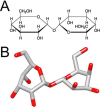
Figure 1. The structure of trehalose
Trehalose is a non-reducing disaccharide composed of two glucose units with a α-1,1-glycosidic linkage. (A) Chemical drawing and (B) three-dimensional structure of trehalose. Red indicates oxygen atoms.
Figure 2
Figure 2. Three proposed theories how trehalose may protect proteins from damage
Figure 3
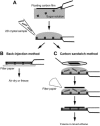
Figure 3. Sugar embedding methods used for the preparation of 2D crystals
(A) A piece of carbon film is floated off mica onto the embedding solution, picked up with an EM grid, and the 2D crystal sample is applied to the opposite side of the grid. (B) In the back-injection method, the grid is simply blotted and then typically allowed to air-dry and cooled down in the electron microscope, but the grid can also be quick-frozen. (C) In the carbon sandwich method, a second, smaller piece of carbon film is placed on the sample using a wire loop. Excess solution is blotted away with a filter paper and the grid quick-frozen in liquid ethane.
Figure 4
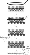
Figure 4. Trehalose embedding used for 2D crystals grown on lipid monolayers
The lipid monolayer with the attached 2D crystal is transferred with a wire loop onto the carbon film on an EM grid, and the trehalose solution is applied to the opposite side of the grid. Excess solution is blotted away with a filter paper and the grid quick-frozen in liquid ethane.
Figure 5
Figure 5. Structure analysis of trehalose-embedded SbpA monolayer 2D crystals
(A) Image of a trehalose-embedded SbpA 2D crystal grown on a lipid monolayer. Scale bar is 200 nm. (B) Power spectrum of the image shown in (A). Scale bar is (5 nm)−1. (C) Projection map of SbpA at 7 Å resolution. A unit cell with lattice constants a = b = 13.3 nm is outlined in black.
Figure 6
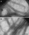
Figure 6. Collagen type 1 fibers prepared by different staining methods
(A) Image of collagen fibers that were spread across a holey carbon film, negatively stained with a trehalose-containing ammonium molybdate solution and air-dried. (B) Image of collagen fibers that were quick-frozen in a saturated ammonium molybdate solution. Scale bar is 300 nm. Figure adapted from Harris, 2008.
Figure 7
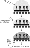
Figure 7. Cryo-negative staining of Affinity Grid preparations
After incubating the Affinity Grid with sample, excess solution is removed with a Hamilton syringe and trehalose-containing staining solution is added to the grid. The grid is then blotted with filter paper and quick-frozen in liquid ethane.
Figure 8
Figure 8. Structure analysis of the Notch extracellular domain (NECD) using a cryo-negatively stained Affinity Grid preparation
The left panel shows a raw image with the white circles indicating individual particles. The scale bar is 25 nm. The right panels show 3D reconstructions of the NECD in three different conformations. Figure adapted from Kelly et al., 2010c.

Similar articles

Cited by

References

    1. Abe K, Tani K, Nishizawa T, Fujiyoshi Y. Inter-subunit interaction of gastric H+,K+-ATPase prevents reverse reaction of the transport cycle. EMBO J. 2009;28:1637–1643. - PMC - PubMed
    1. Abe K, Tani K, Fujiyoshi Y. Conformational rearrangement of gastric H+,K+- ATPase induced by an acid suppressant. Nat. Commun. 2011;2:155. - PMC - PubMed
    1. Abeyrathne PD, Chami M, Pantelic RS, Goldie KN, Stahlberg H. Preparation of 2D crystals of membrane proteins for high-resolution electron crystallography data collection. Methods Enzymol. 2010;481:25–43. - PubMed
    1. Adrian M, Dubochet J, Lepault J, McDowall AW. Cryo-electron microscopy of viruses. Nature. 1984;308:32–36. - PubMed
    1. Adrian M, Dubochet J, Fuller SD, Harris JR. Cryo-negative staining. Micron. 1998;29:145–160. - PubMed

Publication types

MeSH terms

LinkOut - more resources