Comparative Study
. 2011 Sep;134(Pt 9):2548-64.
doi: 10.1093/brain/awr160. Epub 2011 Jul 12.

A comparative clinical, pathological, biochemical and genetic study of fused in sarcoma proteinopathies

Affiliations
Comparative Study

A comparative clinical, pathological, biochemical and genetic study of fused in sarcoma proteinopathies

Tammaryn Lashley et al. Brain. 2011 Sep.

Abstract

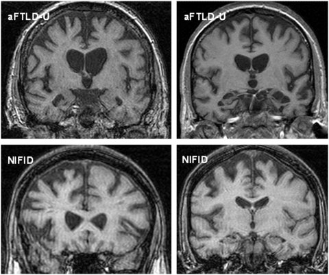
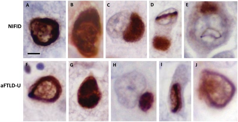
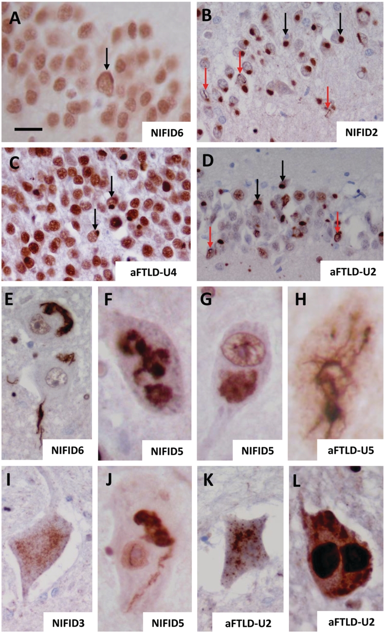
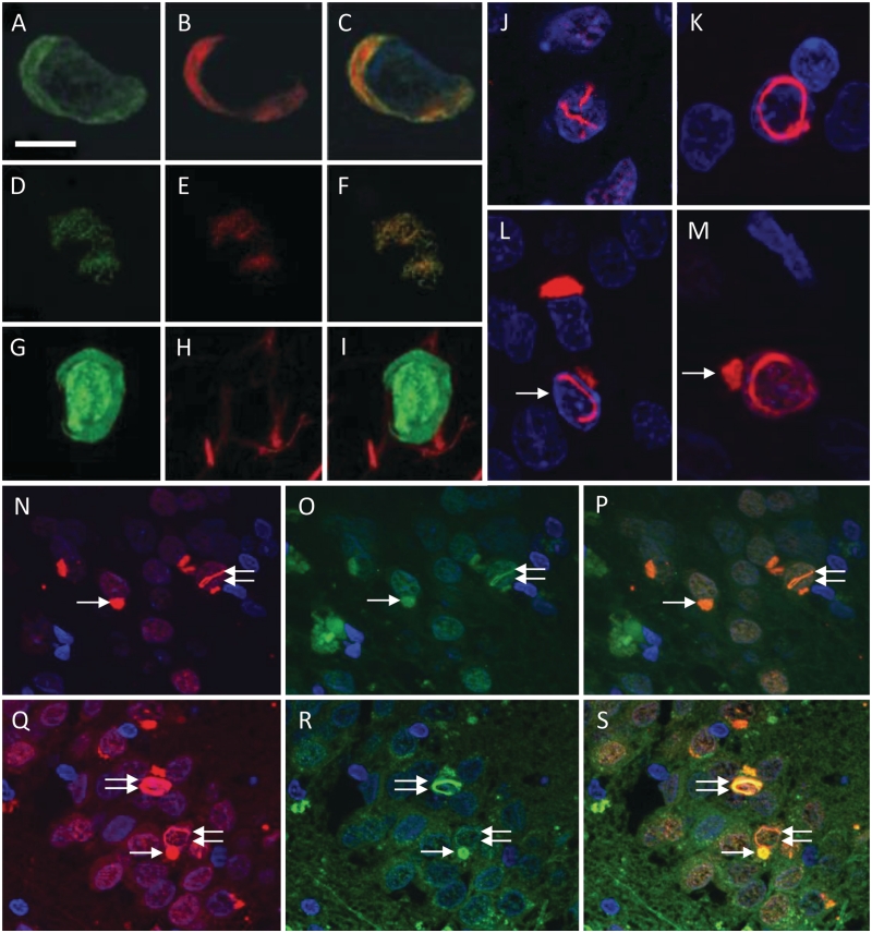
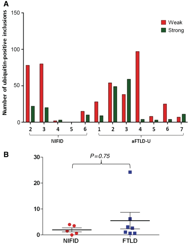
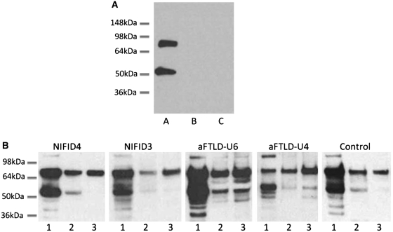
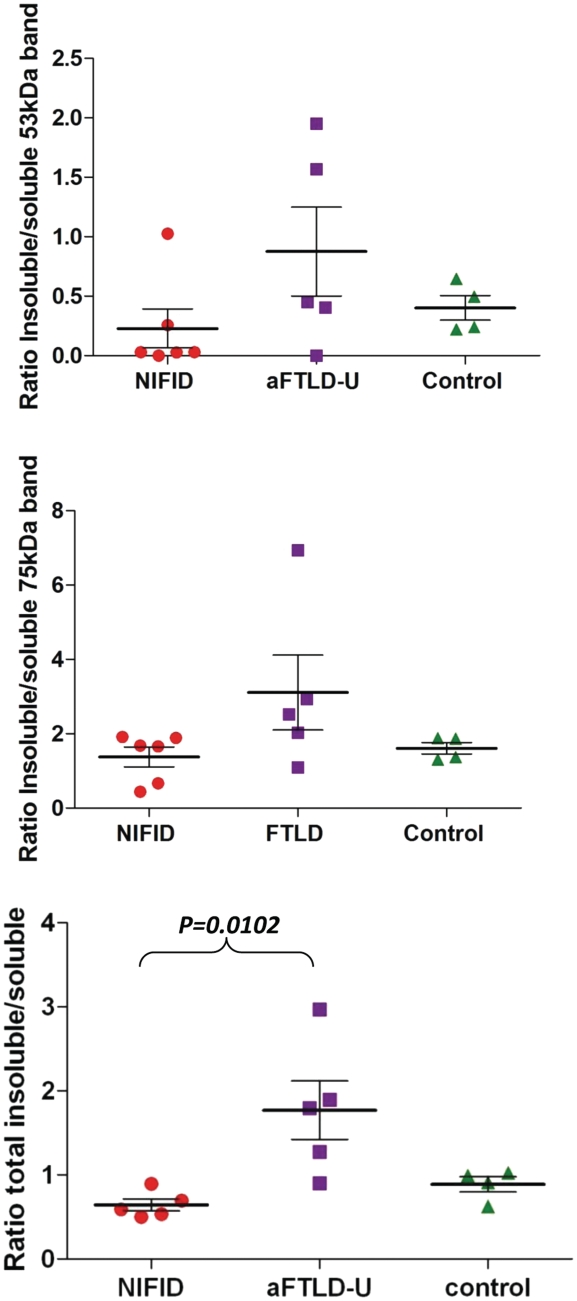
Neuronal intermediate filament inclusion disease and atypical frontotemporal lobar degeneration are rare diseases characterized by ubiquitin-positive inclusions lacking transactive response DNA-binding protein-43 and tau. Recently, mutations in the fused in sarcoma gene have been shown to cause familial amyotrophic lateral sclerosis and fused in sarcoma-positive neuronal inclusions have subsequently been demonstrated in neuronal intermediate filament inclusion disease and atypical frontotemporal lobar degeneration with ubiquitinated inclusions. Here we provide clinical, imaging, morphological findings, as well as genetic and biochemical data in 14 fused in sarcoma proteinopathy cases. In this cohort, the age of onset was variable but included cases of young-onset disease. Patients with atypical frontotemporal lobar degeneration with ubiquitinated inclusions all presented with behavioural variant frontotemporal dementia, while the clinical presentation in neuronal intermediate filament inclusion disease was more heterogeneous, including cases with motor neuron disease and extrapyramidal syndromes. Neuroimaging revealed atrophy of the frontal and anterior temporal lobes as well as the caudate in the cases with atypical frontotemporal lobar degeneration with ubiquitinated inclusions, but was more heterogeneous in the cases with neuronal intermediate filament inclusion disease, often being normal to visual inspection early on in the disease. The distribution and severity of fused in sarcoma-positive neuronal cytoplasmic inclusions, neuronal intranuclear inclusions and neurites were recorded and fused in sarcoma was biochemically analysed in both subgroups. Fused in sarcoma-positive neuronal cytoplasmic and intranuclear inclusions were found in the hippocampal granule cell layer in variable numbers. Cortical fused in sarcoma-positive neuronal cytoplasmic inclusions were often 'Pick body-like' in neuronal intermediate filament inclusion disease, and annular and crescent-shaped inclusions were seen in both conditions. Motor neurons contained variable numbers of compact, granular or skein-like cytoplasmic inclusions in all fused in sarcoma-positive cases in which brainstem and spinal cord motor neurons were available for study (five and four cases, respectively). No fused in sarcoma mutations were found in any cases. Biochemically, two major fused in sarcoma species were found and shown to be more insoluble in the atypical frontotemporal lobar degeneration with ubiquitinated inclusions subgroup compared with neuronal intermediate filament inclusion disease. There is considerable overlap and also significant differences in fused in sarcoma-positive pathology between the two subgroups, suggesting they may represent a spectrum of the same disease. The co-existence of fused in sarcoma-positive inclusions in both motor neurons and extramotor cerebral structures is a characteristic finding in sporadic fused in sarcoma proteinopathies, indicating a multisystem disorder.


Figures

Figure 1
Figure 1
Representative magnetic resonance coronal T1 images of cases with atypical FTLD-U (Top left aFTLD-U6; Top right aFTLD-U3) and NIFID (both images from NIFID1). Top left: the magnetic resonance image shows asymmetrical right greater than left frontotemporal atrophy; Top right: the magnetic resonance image shows asymmetrical left greater than right frontotemporal atrophy. The images on the bottom row are from the same patient and show asymmetrical fronto-insular atrophy.
Figure 2
Figure 2
FUS pathology in NIFID and atypical FTLD-U. Different inclusion types were seen in both NIFID and atypical FTLD-U subgroups. Neurons containing crescent/annular-shaped neuronal cytoplasmic inclusions (A, B, F and G). The strong nuclear staining is retained in B and G, while it is decreased in A and F. Pick body-like inclusions were seen in the NIFID cases (C), whereas smaller rounded neuronal ‘bean shaped' cytoplasmic inclusions were found in atypical FTLD-U (H). Neuronal intranuclear rod shaped (D and I) and vermiform inclusions (E and J) were seen in both NIFID and atypical FTLD-U. Scale bar = 5 µm (A–J).
Figure 3
Figure 3
FUS pathology in the hippocampus (A–D), 12th cranial nerve nucleus (E–H) and spinal cord (I–L) in NIFID and atypical FTLD-U. FUS pathology was variable in the hippocampus. Apart from Patient NIFID5 where it was absent, it ranged from mild to severe in both NIFID (A: mild, B: severe) and atypical FTLD-U (C: mild, D: severe). Neuronal cytoplasmic inclusions (black arrows) and neuronal intranuclear inclusions (red arrows) were present in the granule cell layer of the dentate fascia in both NIFID and atypical FTLD-U. Different FUS-positive inclusions in motor neurons of the 12th cranial nerve nucleus (E–H) and spinal cord (I–L), which included filamentous (E and J), dot-like/granular (I and K), large globular (F, G and L) and skein-like neuronal cytoplasmic inclusions (H) inclusions. Occasional motor neurons also contained intranuclear inclusions (J). Scale bar = 20 µm (A and C); 50 µm (B and D); and 5 µm (E–L).
Figure 4
Figure 4
FUS immunofluorescence studies in NIFID and atypical FTLD-U. Double immunofluorescence with FUS (A and D) and p62 (B and E; combined images C and F). Co-localization of FUS and p62 shown in a crescent-shaped inclusion of a frontal cortical neuron (A–C) and in a skein-like inclusion of a motor neuron of the 12th nerve nucleus (D–F) in NIFID. Double staining with antibodies to FUS and α-internexin (G–I) shows absence of α-internexin (H) in a FUS-positive cortical neuronal cytoplasmic inclusion (G) in NIFID. FUS-positive neuronal intranuclear inclusions demonstrating a number of morphological phenotypes; the image on J shows a ‘double’ intranuclear inclusion while the image on K shows a circular neuronal intranuclear inclusion. Some of the neurons on L and M contained both cytoplasmic and nuclear inclusions (arrows) [(J–M) Granule cells of the dentate fascia, Case NIFID6]. Strong colocalization of FUS (N and Q) and ubiquitin staining (O and R; combined images P and S) in both neuronal cytoplasmic (arrow) and neuronal intranuclear inclusions (double arrow) is demonstrated in granule cells of the dentate fascia in NIFID2 (N–P) and in Patient aFTLD-U4 (Q–S). Scale bar = 10 µm (A–C and G–I); 20 µm (J–M); and 50 µm (N–S).
Figure 5
Figure 5
Semi-quantitative analysis of ubiquitin-positive inclusions in NIFID and atypical FTLD-U. In both NIFID and atypical FTLD-U, the intensity of ubiquitin immunoreactivity of the neuronal inclusions varied from weak/moderate to strong, mirroring variation in FUS immunoreactivity. The number of weakly/moderately and strongly stained ubiquitin-positive inclusions were counted in the granule cell layer of the dentate fascia in each case. (A) Distribution in the NIFID and atypical FTLD-U cases. (B) A ratio of the weakly/moderately and strongly stained inclusions was determined in each case. The mean of the ratios did not differ in the two disease groups (Mann–Whitney U test; P = 0.75).
Figure 6
Figure 6
Biochemical analysis of FUS. (A) Immunoblotting with a FUS antibody demonstrates two major bands at ∼75 and ∼53 kDa in NIFID4 (lane A). Control experiments by omitting the primary antibody (lane B) and using a FUS antibody blocked with the corresponding synthetic peptide (lane C) show no visible bands on the western blot. (B) Proteins were sequentially extracted from NIFID, atypical FTLD-U and control cases. High salt (lane 1), SDS (lane 2) and urea (lane 3) fractions were separated and immunoblotted with an anti-FUS antibody (NB100-565). All cases showed strong ∼75 and ∼53 kDa bands in the soluble fraction and a higher band at ∼120 kDa was also observed mainly in the soluble fraction. All fractions and all cases contained the higher ∼75 kDa band, but the amount of ∼53 kDa FUS species varied between cases with a strong band visible in the atypical FTLD-U cases but generally weaker bands in the NIFID and control cases.
Figure 7
Figure 7
Ratio of insoluble to soluble FUS. The band intensities of FUS in insoluble (SDS and urea soluble) and soluble (PBS soluble) fractions were analysed and a ratio calculated in each case. Bands visible at both ∼53 and ∼75 kDa were analysed individually and combined to calculate the ratios of total FUS. The ratios are illustrated on the plot with each point representing a case and the solid line representing the mean. Although there is some overlap between the two disease types, the atypical FTLD-U group showed significantly higher ratios compared with both the NIFID and control group when total FUS was analysed (one way Kruskal–Wallis test; P = 0.01).

Comment in

References

    1. Aman P, Panagopoulos I, Lassen C, Fioretos T, Mencinger M, Toresson H, et al. Expression patterns of the human sarcoma-associated genes FUS and EWS and the genomic structure of FUS. Genomics. 1996;37:1–8. - PubMed
    1. Andersson MK, Stahlberg A, Arvidsson Y, Olofsson A, Semb H, Stenman G, et al. The multifunctional FUS, EWS and TAF15 proto-oncoproteins show cell type-specific expression patterns and involvement in cell spreading and stress response. BMC Cell Biol. 2008;9:37. - PMC - PubMed
    1. Arai T, Hasegawa M, Akiyama H, Ikeda K, Nonaka T, Mori H, et al. TDP-43 is a component of ubiquitin-positive tau-negative inclusions in frontotemporal lobar degeneration and amyotrophic lateral sclerosis. Biochem Biophys Res Commun. 2006;351:602–11. - PubMed
    1. Armstrong RA, Gearing M, Bigio EH, Cruz-Sanchez FF, Duyckaerts C, Mackenzie IR, et al. The spectrum and severity of FUS-immunoreactive inclusions in the frontal and temporal lobes of ten cases of neuronal intermediate filament inclusion disease. Acta Neuropathol. 2011;121:219–28. - PMC - PubMed
    1. Baechtold H, Kuroda M, Sok J, Ron D, Lopez BS, Akhmedov AT. Human 75-kDa DNA-pairing protein is identical to the pro-oncoprotein TLS/FUS and is able to promote D-loop formation. J Biol Chem. 1999;274:34337–42. - PubMed

Publication types

MeSH terms