. 2012 Feb 7;9(67):261-71.
doi: 10.1098/rsif.2011.0357. Epub 2011 Jul 13.

Enhanced mesenchymal stromal cell recruitment via natural killer cells by incorporation of inflammatory signals in biomaterials

Affiliations

Enhanced mesenchymal stromal cell recruitment via natural killer cells by incorporation of inflammatory signals in biomaterials

Catarina R Almeida et al. J R Soc Interface. .

Abstract

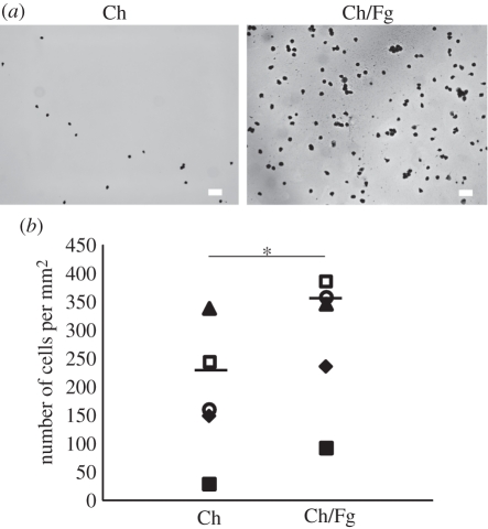
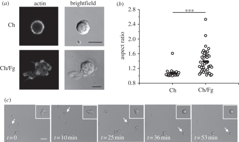
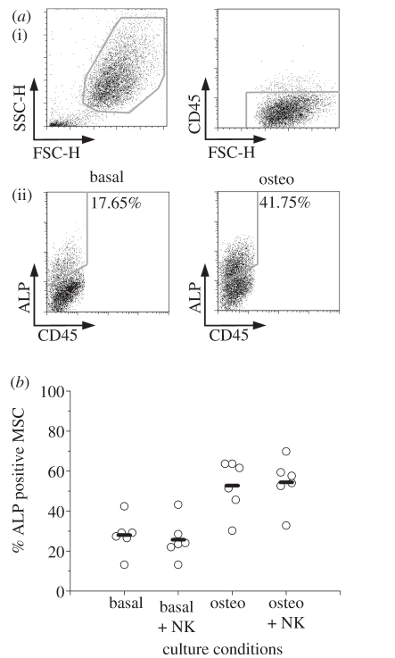
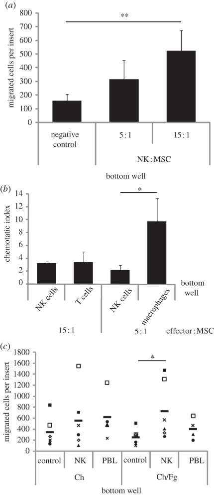
An exacerbated inflammatory response questions biomaterial biocompatibility, but on the other hand, inflammation has a central role in the regulation of tissue regeneration. Therefore, it may be argued that an 'ideal' inflammatory response is crucial to achieve efficient tissue repair/regeneration. Natural killer (NK) cells, being one of the first populations arriving at an injury site, can have an important role in regulating bone repair/regeneration, particularly through interactions with mesenchymal stem/stromal cells (MSCs). Here, we studied how biomaterials designed to incorporate inflammatory signals affected NK cell behaviour and NK cell-MSC interactions. Adsorption of the pro-inflammatory molecule fibrinogen (Fg) to chitosan films led to a 1.5-fold increase in adhesion of peripheral blood human NK cells, without an increase in cytokine secretion. Most importantly, it was found that NK cells are capable of stimulating a threefold increase in human bone marrow MSC invasion, a key event taking place in tissue repair, but did not affect the expression of the differentiation marker alkaline phosphatase (ALP). Of significant importance, this NK cell-mediated MSC recruitment was modulated by Fg adsorption. Designing novel biomaterials leading to rational modulation of the inflammatory response is proposed as an alternative to current bone regeneration strategies.


Figures

Figure 1.
Figure 1.
Increased NK cell adhesion to Ch films with adsorbed Fg. NK cells were incubated for 1 h in Ch films without (Ch) and with adsorbed Fg (Ch/Fg), washed, fixed and stained. Cells were imaged in an inverted microscope and counted. (a) Representative images of cells adherent to films. Scale bar, 40 µm. (b) Summary of data obtained in five independent experiments. Each symbol represents the average number of cells in one experiment with three replicas. Bars show the average of the different experiments. Data are statistically significant (*p < 0.05; Wilcoxon test).
Figure 2.
Figure 2.
NK cells show a migratory phenotype in the presence of adsorbed Fg. (a) NK cells were incubated for 1 h in Ch films (Ch) or Ch films with adsorbed Fg (Ch/Fg), before staining for actin. Scale bar, 5 µm. (b) The aspect ratio, measured from images obtained after actin staining. Circles represent individual cells, bars represent the mean. Data shown were collected in five different experiments performed with NK cells from different donors. Statistical significance was determined with Mann–Whitney test (***p < 0.001). (c) Time-lapse movies were obtained by imaging freshly isolated NK cells on Ch/Fg films every 1 min for 2 h at 37°C/5%CO2, after 1 h of incubation. Snapshots taken at different time points of a representative movie are shown, with a white arrow pointing to an NK cell that exhibited different behaviours: migratory, from t = 0 to t = 35 min, and stopped from t = 36 min to t = 53 min. Scale bar, 20 µm.
Figure 3.
Figure 3.
Production of IFN-γ by NK cells is stimulated by MSCs but is not affected by Fg adsorbed to Ch films. (a) NK cells were incubated alone (i), in the presence of MSCs at a 1 : 1 ratio (ii) or with PMA + ionomycin (iii) in tissue culture polystyrene (TCPS), Ch or Ch/Fg substrates and stained for surface CD56 and intracellular IFN-γ and analysed by flow cytometry. NK cells were gated on the basis of size and CD56 positive staining. Gates were drawn from isotype control stainings. Plots show data from one experiment out of three. Percentage of cells in each quadrant is indicated. (b) The average ± s.e.m. percentage of IFN-γ positive NK cells was plotted for different substrates (n = 3). There were no significant differences between substrates (Wilcoxon test). Black bars, basal; dark grey bars, MSC; light grey bars, PMA.
Figure 4.
Figure 4.
NK cells do not affect ALP expression by MSCs. MSCs were cultured in the absence or presence of osteogenic stimuli (basal or osteo) and in the absence or presence of NK cells, at a 5 : 1 NK:MSC ratio for 7 days, and stained for surface ALP and CD45. (a) (i) Dot plots show how MSCs were distinguished on the basis of size and negative expression of CD45. (ii) Osteogenic conditions lead to an increase in the percentage of MSCs expressing surface ALP. Numbers indicate the percentage of cells within the drawn gates (ALP positive). (b) Summary of data obtained in six independent experiments. Each circle shows the percentage of ALP positive MSCs in one experiment. Bars show the average of different experiments. Differences between basal and basal + NK or between osteo and osteo + NK conditions are not statistically significant (Wilcoxon test).
Figure 5.
Figure 5.
Increased MSC invasion through Matrigel in the presence of NK cells. MSC invasion through filters with 8 µm pores and coated with Matrigel was determined after 24 h incubation. The number of migrated cells per insert was estimated upon counting 10 fields of view per sample (200×). (a) The lower compartment of the invasion chamber was filled with serum-free medium, as a negative control, or with serum-free medium with NK cells at an NK : MSC ratio of 5 : 1 or 15 : 1. Graph shows the mean ± s.e.m. of five to 10 independent experiments. (b) MSCs invasion in the presence of NK cells, T cells or macrophages. Chemotatic index refers to the number of migrated MSCs in the presence of the indicated cell type, divided by the number of migrated MSCs in the negative control. Graph shows the mean ± s.e.m. of experiments performed with at least four independent blood donors. (*p < 0.05; Mann–Whitney test). (c) The lower compartment of the chamber was filled with serum-free medium, as a negative control, with serum-free medium with NK cells, or with PBLs at an effector : MSC ratio of 15 : 1, either seeded on Ch or Ch/Fg films. Each symbol corresponds to data from one experiment. Different symbols correspond to data obtained with cells from different donors. Bars show the average of the different experiments. (*p < 0.05, **p < 0.01; Wilcoxon test).

References

    1. Mountziaris P. M., Mikos A. G. 2008. Modulation of the inflammatory response for enhanced bone tissue regeneration. Tissue Eng. Part B Rev. 14, 179–18610.1089/ten.teb.2008.0038 (doi:10.1089/ten.teb.2008.0038) - DOI - DOI - PMC - PubMed
    1. Anderson J. M., Rodriguez A., Chang D. T. 2008. Foreign body reaction to biomaterials. Semin. Immunol. 20, 86–10010.1016/j.smim.2007.11.004 (doi:10.1016/j.smim.2007.11.004) - DOI - DOI - PMC - PubMed
    1. Agaiby A. D., Dyson M. 1999. Immuno-inflammatory cell dynamics during cutaneous wound healing. J. Anat. 195, 531–54210.1046/j.1469-7580.1999.19540531.x (doi:10.1046/j.1469-7580.1999.19540531.x) - DOI - DOI - PMC - PubMed
    1. Vivier E., Tomasello E., Baratin M., Walzer T., Ugolini S. 2008. Functions of natural killer cells. Nat. Immunol. 9, 503–51010.1038/ni1582 (doi:10.1038/ni1582) - DOI - DOI - PubMed
    1. Aggarwal S., Pittenger M. F. 2005. Human mesenchymal stem cells modulate allogeneic immune cell responses. Blood 105, 1815–182210.1182/blood-2004-04-1559 (doi:10.1182/blood-2004-04-1559) - DOI - DOI - PubMed

Publication types

LinkOut - more resources