

Functional imaging of SDHx-related head and neck paragangliomas: comparison of 18F-fluorodihydroxyphenylalanine, 18F-fluorodopamine, 18F-fluoro-2-deoxy-D-glucose PET, 123I-metaiodobenzylguanidine scintigraphy, and 111In-pentetreotide scintigraphy
- PMID: 21752889
- PMCID: PMC3167674
- DOI: 10.1210/jc.2011-0333
Functional imaging of SDHx-related head and neck paragangliomas: comparison of 18F-fluorodihydroxyphenylalanine, 18F-fluorodopamine, 18F-fluoro-2-deoxy-D-glucose PET, 123I-metaiodobenzylguanidine scintigraphy, and 111In-pentetreotide scintigraphy
Abstract
Rationale: Accurate diagnosis of head and neck paragangliomas is often complicated by biochemical silence and lack of catecholamine-associated symptoms, making accurate anatomical and functional imaging techniques essential to the diagnostic process.
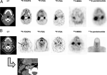
Methods: Ten patients (seven SDHD, three SDHB), with a total of 26 head and neck paragangliomas, were evaluated with anatomical and functional imaging. This study compares five different functional imaging techniques [(18)F-fluorodihydroxyphenylalanine ((18)F-FDOPA) positron emission tomography (PET), (18)F-fluorodopamine ((18)F-FDA) PET/computed tomography (CT), (18)F-fluoro-2-deoxy-D-glucose ((18)F-FDG) PET/CT, (123)I-metaiodobenzylguanidine ((123)I-MIBG) scintigraphy, and (111)In-pentetreotide scintigraphy] in the localization of head and neck paragangliomas.
Results: Prospectively (18)F-FDOPA PET localized 26 of 26 lesions in the 10 patients, CT/magnetic resonance imaging localized 21 of 26 lesions, (18)F-FDG PET/CT localized 20 of 26 lesions, (111)In-pentetreotide scintigraphy localized 16 of 25 lesions, (18)F-FDA PET/CT localized 12 of 26 lesions, and (123)I-MIBG scintigraphy localized eight of 26 lesions. Differences in imaging efficacy related to genetic phenotype, even in the present small sample size, included the negativity of (18)F-FDA PET/CT and (123)I-MIBG scintigraphy in patients with SDHB mutations and the accuracy of (18)F-FDG PET/CT in all patients with SDHD mutations, as compared with the accuracy of (18)F-FDG PET/CT in only one patient with an SDHB mutation.
Conclusion: Overall, (18)F-FDOPA PET proved to be the most efficacious functional imaging modality in the localization of SDHx-related head and neck paragangliomas and may be a potential first-line functional imaging agent for the localization of these tumors.
Figures
References
-
- Gujrathi CS, Donald PJ. 2005. Current trends in the diagnosis and management of head and neck paragangliomas. Curr Opin Otolaryngol Head Neck Surg 13:339–342 - PubMed
-
- Erickson D, Kudva YC, Ebersold MJ, Thompson GB, Grant CS, van Heerden JA, Young WF., Jr 2001. Benign paragangliomas: clinical presentation and treatment outcomes in 236 patients. J Clin Endocrinol Metab 86:5210–5216 - PubMed
-
- Baysal BE, Willett-Brozick JE, Lawrence EC, Drovdlic CM, Savul SA, McLeod DR, Yee HA, Brackmann DE, Slattery WH, 3rd, Myers EN, Ferrell RE, Rubinstein WS. 2002. Prevalence of SDHB, SDHC, and SDHD germline mutations in clinic patients with head and neck paragangliomas. J Med Genet 39:178–183 - PMC - PubMed
-
- Neumann HP, Pawlu C, Peczkowska M, Bausch B, McWhinney SR, Muresan M, Buchta M, Franke G, Klisch J, Bley TA, Hoegerle S, Boedeker CC, Opocher G, Schipper J, Januszewicz A, Eng C. 2004. Distinct clinical features of paraganglioma syndromes associated with SDHB and SDHD gene mutations. JAMA 292:943–951 - PubMed
-
- Pacak K, Eisenhofer G, Goldstein DS. 2004. Functional imaging of endocrine tumors: role of positron emission tomography. Endocr Rev 25:568–580 - PubMed
Publication types
MeSH terms
Substances
Grants and funding
LinkOut - more resources
Full Text Sources
Medical
