. 2011 Sep;85(18):9614-22.
doi: 10.1128/JVI.00480-11. Epub 2011 Jul 13.

The leader protein of cardioviruses inhibits stress granule assembly

Affiliations

The leader protein of cardioviruses inhibits stress granule assembly

Fabian Borghese et al. J Virol. 2011 Sep.

Abstract

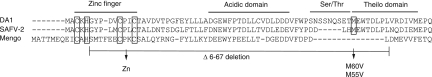
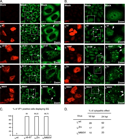
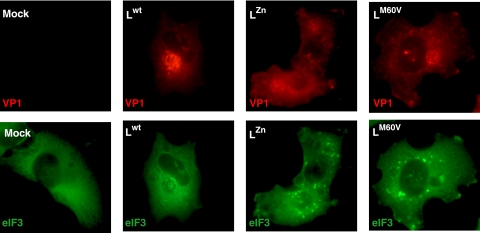
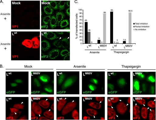
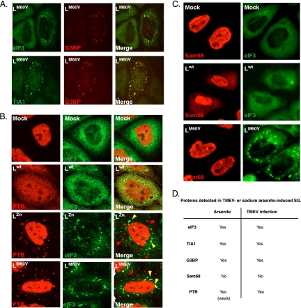
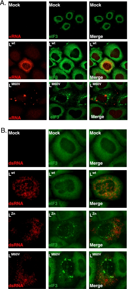
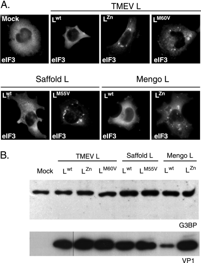
Stress granules (SG) are cytoplasmic aggregates of stalled translation preinitiation complexes that form in cells exposed to various environmental stresses. Here, we show that stress granules assemble in cells infected with Theiler's murine encephalomyelitis virus (TMEV) mutants carrying alterations in the leader (L) protein, but not in cells infected with wild-type TMEV. Stress granules also formed in STAT1-deficient cells, suggesting that SG formation was not a consequence of increased type I interferon (IFN) production when cells were infected with the mutant virus. Ectopic expression of the wild-type L protein was sufficient to inhibit stress granule formation induced by sodium arsenite or thapsigargin treatment. In conclusion, TMEV infection induces stress granule assembly, but this process is inhibited by the L protein. Unlike poliovirus-induced stress granules, TMEV-induced stress granules did not contain the nuclear protein Sam68 but contained polypyrimidine tract binding protein (PTB), an internal ribosome entry site (IRES)-interacting protein. Moreover, G3BP was not degraded and was found in SG after TMEV infection, suggesting that SG content could be virus specific. Despite the colocalization of PTB with SG and the known interaction of PTB with viral RNA, in situ hybridization and immunofluorescence assays failed to detect viral RNA trapped in infection-induced SG. Recombinant Theiler's viruses expressing the L protein of Saffold virus 2 (SAFV-2), a closely related human theilovirus, or the L protein of mengovirus, an encephalomyocarditis virus (EMCV) strain, also inhibited infection-induced stress granule assembly, suggesting that stress granule antagonism is a common feature of cardiovirus L proteins.


Figures

Fig. 1.
Fig. 1.
Alignment of L protein sequences used in this work. Shown are the sequences of L proteins from TMEV (strain DA1), SAFV-2, and mengovirus. Protein domains are indicated. Cysteine and histidine residues forming the Zn finger, and the M60/M55 residue of the Theilo domain, are framed. L mutations are indicated under the alignment.
Fig. 2.
Fig. 2.
TMEV infection induces stress granule assembly. (A) HeLa cells were infected with 10 PFU/cell of wild-type TMEV (Lwt) or with LΔ6–67, LZn, or LM60V mutant virus. Twelve hours postinfection cells were fixed, coimmunostained for the VP1 capsid viral antigen (red) and eIF3 (green), and examined by confocal microscopy. Arrowheads point to infected cells (i.e., VP1 positive). The right column (zoom) shows a higher-magnification view of the region framed in the middle column. Note that some VP1-negative cells also display SG in the wells infected with the L-mutant viruses. This is probably due to the fact that VP1 had not reached a detectable level at the time when the cells were fixed. (B) Same experiment as in panel A but with immunostaining of VP1 (red) and TIA-1 (green). (C) Histogram showing the percentage (mean of two independent experiments) of VP1-positive cells displaying SG according to the virus used. Note that cells presenting CPE were excluded from the counts. (D) Percentages (means of two independent experiments) of cytopathic effect observed in HeLa cells infected with the wild-type and mutant viruses at 16 and 24 h postinfection.
Fig. 3.
Fig. 3.
Inhibition of SG assembly is independent of L inhibition of interferon production. U3A cells were infected with 5 PFU/cell of wild-type TMEV (Lwt) or with LZn and LM60V mutant viruses. At 8 hours postinfection, cells were processed for VP1 and eIF3 coimmunolabeling and examined by fluorescence microscopy. SG appear as bright spots of eIF3 staining in the cytoplasm.
Fig. 4.
Fig. 4.
Ectopic expression of L is sufficient to inhibit arsenite- or thapsigargin-induced SG assembly. (A) HeLa cells were infected with a wild-type TMEV (Lwt) or with L-mutant viruses (not shown). Twelve hours postinfection, cells were treated with 0.5 mM sodium arsenite for 45 min, fixed, and processed for coimmunolabeling of VP1 and eIF3 (confocal microscopy images). (B) HeLa cells were transfected with bicistronic constructs expressing Lwt and eGFP or LM60V and eGFP. Sixteen hours posttransfection cells were mock treated or treated with 0.5 mM sodium arsenite for 45 min or with 15 μM thapsigargin for 50 min, fixed, and processed for eIF3 immunolabeling. White arrowheads indicate transfected cells. Note that cells transfected with the Lwt-IRES-eGFP construct had a much lower eGFP fluorescence level because Lwt expression represses eGFP expression (30). Moreover, eIF3 exhibited a partially nuclear localization in many cells expressing Lwt, in agreement with the reported effect of this protein on nucleocytoplasmic transport. (C) Histogram showing the percentages of transfected (eGFP-positive) cells displaying total or partial inhibition or no inhibition of arsenite- and thapsigargin-induced SG. In view of ectopically expressed L protein toxicity, only cells with unaltered morphology were taken into account.
Fig. 5.
Fig. 5.
PTB, but not Sam68, partially colocalizes with TMEV infection-induced SG. (A) Confocal microscopy images showing coimmunostaining of eIF3 and G3BP or TIA-1 and G3BP in HeLa cells infected for 16 h (10 PFU/cell) with the LM60V mutant virus. (B) Confocal microscopy images showing coimmunostaining of eIF3 and PTB in HeLa cells infected for 12 h (10 PFU/cell) with wild-type TMEV or with LZn and LM60V mutant viruses. Yellow arrowheads indicate colocalization between PTB and stress granules. Red arrowheads indicate cytoplasmic PTB foci in which no eIF3 was detected. (C) Double fluorescence microscopy images showing representative U3A cells coimmunostained for Sam68 and eIF3, 8 h after infection with 5 PFU/cell of wild-type or L-mutant (LM60V) TMEV. (D) Detected stress granule markers associated with arsenite-induced SG or TMEV infection-induced SG.
Fig. 6.
Fig. 6.
Viral RNA is not detected in TMEV-induced SG. (A) In situ hybridization (ISH) to detect positive-stranded viral RNA coupled to immunolabeling of eIF3. HeLa cells were mock infected or infected with wild-type or LM60V TMEV at 10 PFU per cell. After 12 h of infection, cells were fixed and processed for ISH (red) and immunolabeling of eIF3 (green) (confocal microscopy images). (B) Coimmunolabeling of the viral double-stranded RNA (dsRNA; detected with the K1 antibody; red) and eIF3 (green). Experimental conditions were the same as in Fig. 2. (conventional microscopy images).
Fig. 7.
Fig. 7.
Inhibition of SG assembly is a common activity of cardiovirus L protein. (A) U3A cells were infected in parallel with a wild-type TMEV or with TMEV recombinants expressing TMEV LZn, TMEV LM60V, SAFV-2 Lwt, SAFV-2 LM55V, mengovirus Lwt, or mengovirus LZn. At 8 hours postinfection cells were fixed and processed for coimmunolabeling of VP1 (not shown) and eIF3. Each picture shows the distribution of eIF3 in a VP1-positive cell (not shown), except for the control, which was VP1 negative. (B) Western blot detection of G3BP in extracts from U3A cells infected as in panel A. No sign of G3BP cleavage or decrease in band intensity was observed when comparing wild-type and L-mutant viruses. Note that lower VP1 expression in SPA24 (Lwt mengovirus)-infected cells was noticed previously (23) and is thought to result from an altered synchronism between viral replication and L activity, which can indirectly impair viral replication.

Similar articles

Cited by

References

    1. Agol V. I., Gmyl A. P. 2010. Viral security proteins: counteracting host defences. Nat. Rev. Microbiol. 8:867–878 - PMC - PubMed
    1. Anderson P., Kedersha N. 2009. RNA granules: post-transcriptional and epigenetic modulators of gene expression. Nat. Rev. Mol. Cell Biol. 10:430–436 - PubMed
    1. Anderson P., Kedersha N. 2009. Stress granules. Curr. Biol. 19:R397–R398 - PubMed
    1. Bardina M. V., et al. 2009. Mengovirus-induced rearrangement of the nuclear pore complex: hijacking cellular phosphorylation machinery. J. Virol. 83:3150–3161 - PMC - PubMed
    1. Brahic M., Bureau J. F., Michiels T. 2005. The genetics of the persistent infection and demyelinating disease caused by Theiler's virus. Annu. Rev. Microbiol. 59:279–298 - PubMed

Publication types