. 2011 Oct;85(19):10269-78.
doi: 10.1128/JVI.05273-11. Epub 2011 Jul 13.

A dual strategy for the suppression of host antiviral silencing: two distinct suppressors for viral replication and viral movement encoded by potato virus M

Affiliations

A dual strategy for the suppression of host antiviral silencing: two distinct suppressors for viral replication and viral movement encoded by potato virus M

Hiroko Senshu et al. J Virol. 2011 Oct.

Abstract

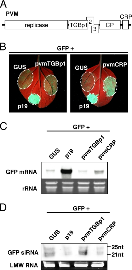
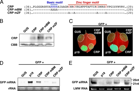
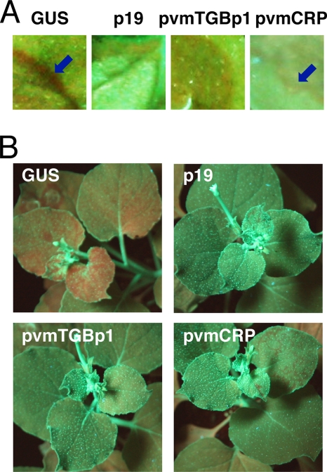
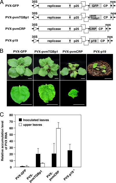
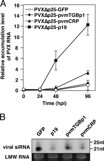
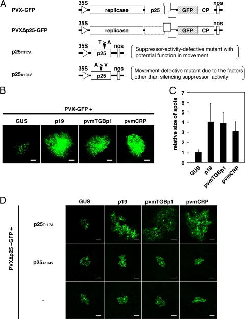
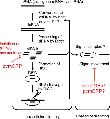
Viruses encode RNA silencing suppressors to counteract host antiviral silencing. In this study, we analyzed the suppressors encoded by potato virus M (PVM), a member of the genus Carlavirus. In the conventional green fluorescent protein transient coexpression assay, the cysteine-rich protein (CRP) of PVM inhibited both local and systemic silencing, whereas the triple gene block protein 1 (TGBp1) showed suppressor activity only on systemic silencing. Furthermore, to elucidate the roles of these two suppressors during an active viral infection, we performed PVX vector-based assays and viral movement complementation assays. CRP increased the accumulation of viral RNA at the single-cell level and also enhanced viral cell-to-cell movement by inhibiting RNA silencing. However, TGBp1 facilitated viral movement but did not affect viral accumulation in protoplasts. These data suggest that CRP inhibits RNA silencing primarily at the viral replication step, whereas TGBp1 is a suppressor that acts at the viral movement step. Thus, our findings demonstrate a sophisticated viral infection strategy that suppresses host antiviral silencing at two different steps via two mechanistically distinct suppressors. This study is also the first report of the RNA silencing suppressor in the genus Carlavirus.


Figures

Fig. 1.
Fig. 1.
Suppressor activity of pvmTGBp1 and pvmCRP on sense transgene-induced local RNA silencing. (A) Schematic representation of the genome of potato virus M showing ORFs. (B) GFP fluorescence images of N. benthamiana leaves infiltrated with Agrobacterium mixtures containing a vector expressing GFP and GUS (left upper patches of each panel), p19 (left lower patches), pvmTGBp1 (right patch of left panel), or pvmCRP (right patch of right panel). Photographs were taken under UV light at 4 dpi. (C and D) Northern blot analysis of GFP mRNA (C) and siRNAs (D) extracted from the infiltrated patches shown in panel B. Ethidium bromide-stained rRNA or low-molecular-weight RNA (LMW RNA) is shown below each panel as a loading control. Molecular size markers are indicated on the right.
Fig. 2.
Fig. 2.
Suppressor activity of pvmCRP mutants on sense transgene-induced local RNA silencing. (A) Schematic representation of the site-directed mutagenesis of pvmCRP. The positions of the basic motif (in blue letters) and the zinc-finger motif (in red letters) are shown. The mutants CRPmBM and CRPmZF harbor alanine substitutions in each motif, and the dashes indicate residues identical to the wild type. Numbers in parentheses indicate amino acid residues not shown here. (B) Immunoblot analysis of pvmCRP and its mutants. Total protein was extracted from the leaves infiltrated with Agrobacterium mixtures containing a vector expressing p19 and GUS, tagged CRP, CRPmBM, or CRPmZF at 3 dpi. Coomassie brilliant blue-stained total protein (CBB) is shown as a loading control. (C) GFP fluorescence images of N. benthamiana leaves infiltrated with Agrobacterium mixtures containing a vector expressing GFP and GUS (left upper patches of each panel), p19 (left lower patches), wild-type CRP (right lower patches) CRPmBM (right upper patch of left panel), or CRPmZF (right upper patch of right panel). The photographs were taken under UV light at 4 dpi. (D and E) Northern blot analysis of GFP mRNA (D) and siRNAs (E) extracted from the infiltrated patches shown in panel C. Ethidium bromide-stained rRNA or low-molecular-weight RNA (LMW RNA) is shown below each panel as a loading control. Molecular size markers are indicated on the right.
Fig. 3.
Fig. 3.
Suppressor activity of pvmTGBp1 and pvmCRP on the spread of RNA silencing. (A and B) GFP fluorescence images of infiltrated (A) or systemic (B) leaves of N. benthamiana line 16c infiltrated with Agrobacterium mixtures containing a vector expressing GFP and either GUS, p19, pvmTGBp1, or pvmCRP are shown. Photographs were taken under UV light at 13 dpi. The short-range movement of GFP silencing is represented by the red ring on the border of the patch (blue arrow). Note that the pvmTGBp1-introduced patch shows the disappearance of GFP fluorescence because of local RNA silencing induction, which is unrelated to the red border. Also, the pvmCRP-introduced patch shows GFP fluorescence due to suppression of local RNA silencing, whereas the red border is faintly visible.
Fig. 4.
Fig. 4.
Effects of pvmTGBp1 and pvmCRP on PVX pathogenicity. (A) Schematic representation of recombinant PVX variants. The GFP, pvmTGBp1, pvmCRP, or TBSV p19 sequence is inserted downstream from the coat protein promoter in an infectious PVX cDNA clone. (B) Symptoms of the PVX-GFP, PVX-pvmTGBp1, PVX-pvmCRP, or PVX p19-infected plants. Photographs of whole plants (upper panels) and of systemically infected leaves (lower panels) were taken at 15 dpi. Scale bars, 3 cm. (C) Quantitative real-time PCR (qRT-PCR) analysis of PVX coat protein (CP) RNA accumulation in each recombinant virus-inoculated plant. Total RNA was extracted from inoculated leaves at 5 dpi (▪) or upper leaves at 16 dpi (□). The accumulation levels relative to PVX-GFP were calculated via the second derivative method using ubiquitin transcripts as the internal standard. The error bars represent the standard deviations of three replicates. Note that the upper leaves of the plant inoculated with PVX-p19 are not shown due to necrosis of the entire plant.
Fig. 5.
Fig. 5.
Effects of pvmTGBp1 and pvmCRP on viral replication. (A) PVXΔp25-GFP, PVXΔp25-pvmTGBp1, PVXΔp25-pvmCRP, or PVXΔp25-p19 was inoculated into N. tabacum BY-2 protoplasts, and qRT-PCR analysis of PVX coat protein (CP) RNA was performed with total RNA extracted from the protoplasts at 0, 24, 48, and 96 hpi. The accumulation levels relative to PVX-GFP at 96 hpi were calculated as described in Fig. 4C. The error bars represent the standard deviations of two independent experiments. (B) Northern blot analysis of viral siRNAs extracted from the protoplasts at 96 hpi as shown in panel A. Ethidium bromide-stained low-molecular-weight RNA (LMW RNA) is shown below each panel as a loading control. Molecular size markers are indicated on the right.
Fig. 6.
Fig. 6.
Effects of pvmTGBp1 and pvmCRP on viral cell-to-cell movement. (A) Schematic representation of recombinant PVX variants and mutant derivatives of PVX p25. PVXΔp25-GFP consists of an infectious PVX cDNA clone that expresses GFP from the CP promoter and harbors stop codons and a deletion in the p25 ORF (broken box). For the p25 mutants, p25T117A and p25A104V, the amino acid substitutions are shown above each ORF (solid triangles). (B) Fluorescence microscopic examination of cell clusters expressing GFP in N. benthamiana leaves cointroduced with PVX-GFP and either GUS, p19, pvmTGBp1, or pvmCRP. Photographs were taken at 6 dpi. Scale bars, 500 μm. (C) Quantification of the size of the green fluorescent spots derived from PVX-GFP. Twenty spots, as shown in panel B, were selected randomly from at least three leaves of two independent replicates for each expression construct (GUS, p19, pvmTGBp1, or pvmCRP), and the fluorescent areas were measured using ImageJ software v1.40 (NIH, Bethesda, MD). The relative sizes normalized to GUS are shown. Error bars represent the standard deviations. (D) Cell-to-cell movement analysis of PVX-GFPΔp25 in leaves expressing p25 mutants (none [–], p25T117A, or p25A104V) and a silencing suppressor (GUS as a control, p19, pvmTGBp1, or pvmCRP). Cells expressing GFP were visualized using fluorescence microscopy and photographs were taken at 5 dpi. Scale bars, 50 μm.
Fig. 7.
Fig. 7.
Model of RNA silencing directed against exogenous RNA species and its suppression by viral suppressor proteins. pvmCRP suppresses intracellular silencing and inhibits the accumulation of siRNA. These suppressors also interfere with the spread of silencing, either due to the inhibition of signal siRNA accumulation at the single cell level or due to interference with the signal movement/recipient step in addition to intracellular silencing. pvmTGBp1 prevents the spread of silencing by specifically targeting the signal movement/recipient step.

Similar articles

Cited by

References

    1. Alvarado V., Scholthof H. B. 2009. Plant responses against invasive nucleic acids: RNA silencing and its suppression by plant viral pathogens. Semin. Cell Dev. Biol. 20:1032–1040 - PMC - PubMed
    1. Azevedo J., et al. 2010. Argonaute quenching and global changes in Dicer homeostasis caused by a pathogen-encoded GW repeat protein. Genes Dev. 24:904–915 - PMC - PubMed
    1. Baulcombe D. 2004. RNA silencing in plants. Nature 431:356–363 - PubMed
    1. Baumberger N., Tsai C. H., Lie M., Havecker E., Baulcombe D. C. 2007. The polerovirus silencing suppressor P0 targets Argonaute proteins for degradation. Curr. Biol. 17:1609–1614 - PubMed
    1. Bayne E. H., Rakitina D. V., Morozov S. Y., Baulcombe D. C. 2005. Cell-to-cell movement of potato potexvirus X is dependent on suppression of RNA silencing. Plant J. 44:471–482 - PubMed

Publication types