. 2011 Sep;85(18):9327-33.
doi: 10.1128/JVI.05015-11. Epub 2011 Jul 13.

The structure of barmah forest virus as revealed by cryo-electron microscopy at a 6-angstrom resolution has detailed transmembrane protein architecture and interactions

Affiliations

The structure of barmah forest virus as revealed by cryo-electron microscopy at a 6-angstrom resolution has detailed transmembrane protein architecture and interactions

Victor A Kostyuchenko et al. J Virol. 2011 Sep.

Abstract

Barmah Forest virus (BFV) is a mosquito-borne alphavirus that infects humans. A 6-Å-resolution cryo-electron microscopy three-dimensional structure of BFV exhibits a typical alphavirus organization, with RNA-containing nucleocapsid surrounded by a bilipid membrane anchored with the surface proteins E1 and E2. The map allows details of the transmembrane regions of E1 and E2 to be seen. The C-terminal end of the E2 transmembrane helix binds to the capsid protein. Following the E2 transmembrane helix, a short α-helical endodomain lies on the inner surface of the lipid envelope. The E2 endodomain interacts with E1 transmembrane helix from a neighboring E1-E2 trimeric spike, thereby acting as a spacer and a linker between spikes. In agreement with previous mutagenesis studies, the endodomain plays an important role in recruiting other E1-E2 spikes to the budding site during virus assembly. The E2 endodomain may thus serve as a target for antiviral drug design.


Figures
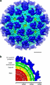
Fig. 1.
Fig. 1.
Overall structure and organization of BFV. (a) The 6-Å-resolution cryo-EM map of BFV low-pass filtered to 10-Å resolution for clarity. Colors are according to radius: green for less than 260 Å, cyan for 260 to 290 Å, and blue for 290 to 350 Å. The scale bar is 100 Å. (b) A quarter of the center section of the BFV cryo-EM map. The densities corresponding to the RNA region, nucleocapsid shell, lipid membrane, and the glycoprotein E1/E2 layers are colored in red, yellow, green, and blue, respectively.
Fig. 2.
Fig. 2.
Overall structure and fitting of E1, E2, and capsid structures into the cryo-EM map of BFV. (a) Arrangement of E1, E2, and capsid in BFV structure. (b) Part of the E1 ectodomain fitted into the cryo-EM density map. (c) The unique glycosylation site on E1 of BFV. The side chain of Asn209 is shown. The density corresponding to the glycan is indicated by a black arrow. (d) Fit of the capsid protein into cryo-EM density and tracing of 10 additional residues, Asn97-Ile106 at the N-terminal end of the protein facing the RNA genome.
Fig. 3.
Fig. 3.
Interactions of E2 transmembrane helix and endodomain. (a) The front (left) and side (right) view of the fit of the transmembrane region of E1 (brown), E2 (light green), and the E2 endodomain (orange) into the cryo-EM map. Capsid protein is colored magenta. Black lines indicate the boundaries of the bilipid membrane. (b) Interaction of the hydrophobic pocket of the capsid protein with the end of the E2 transmembrane helix. The hydrophobic residues are colored in blue. (c) Interaction of E2 endodomain with an E1 transmembrane helix from a neighboring spike (gray ribbon). The distance (8.4 Å) between interacting sites is measured between the centers of the helices. The E1 and E2 transmembrane regions and E2 endodomain are labeled E1TM, E2TM, and E2ed, respectively. Electron densities are shown as gray mesh.
Fig. 4.
Fig. 4.
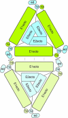
Schematic diagram of two neighboring E1/E2 trimeric spikes. Interactions between ectodomains of E1 on the outside of the plasma membrane (dotted lines) and between E2 endodomains (ed) and E1 transmembrane helices (green TM) (jagged lines) form the links between all E1/E2 trimeric spikes.
Fig. 5.
Fig. 5.
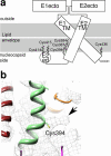
Palmitoylated cysteine residues in E1 and E2. (a) Schematic diagram of the palmitoylation sites on E1and E2 transmembrane helices and E2 endodomain. Palmitoylated residues are indicated by jagged lines. E1 and E2 ectodomains are labeled E1ecto and E2ecto, respectively. (b) Cryo-EM density (black arrow) at Cys394 in the E2 transmembrane helix that corresponds to part of the covalently linked palmitic acid.

References

    1. Caspar D. L., Klug A. 1962. Physical principles in the construction of regular viruses. Cold Spring Harb. Symp. Quant. Biol. 27:1–24 - PubMed
    1. Charollais J., Van Der Goot F. G. 2009. Palmitoylation of membrane proteins (review). Mol. Membr. Biol. 26:55–66 - PubMed
    1. Choi H. K., Lu G., Lee S., Wengler G., Rossmann M. G. 1997. Structure of Semliki Forest virus core protein. Proteins 27:345–359 - PubMed
    1. Combet C., Blanchet C., Geourjon C., Deleage G. 2000. NPS@: network protein sequence analysis. Trends Biochem. Sci. 25:147–150 - PubMed
    1. Dalgarno L., et al. 1984. Characterization of Barmah forest virus: an alphavirus with some unusual properties. Virology 133:416–426 - PubMed

Publication types

Associated data

LinkOut - more resources