. 2011 Sep;85(18):9377-84.
doi: 10.1128/JVI.00178-11. Epub 2011 Jul 13.

The L-coding region of the DA strain of Theiler's murine encephalomyelitis virus causes dysfunction and death of myelin-synthesizing cells

Affiliations

The L-coding region of the DA strain of Theiler's murine encephalomyelitis virus causes dysfunction and death of myelin-synthesizing cells

G D Ghadge et al. J Virol. 2011 Sep.

Abstract

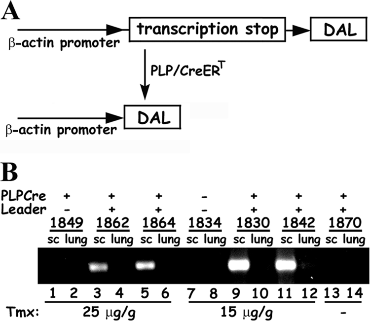
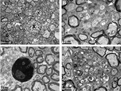
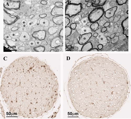
The DA strain and other members of the TO subgroup of Theiler's murine encephalomyelitis virus (TMEV) induce an early transient subclinical neuronal disease followed by a chronic progressive inflammatory demyelination, with persistence of the virus in the central nervous system (CNS) for the life of the mouse. Although TMEV-induced demyelinating disease (TMEV-IDD) is thought to be immune mediated, there is also evidence that supports a role for the virus in directly inducing demyelination. In order to clarify the function of DA virus genes, we generated a transgenic mouse that had tamoxifen-inducible expression of the DA L-coding region in oligodendrocytes (and Schwann cells), a cell type in which the virus is known to persist. Tamoxifen-treated young transgenic mice usually developed an acute progressive fatal paralysis, with abnormalities of the oligodendrocytes and Schwann cells and demyelination, but without significant lymphocytic infiltration; later treatment led to transient weakness with demyelination and persistent expression of the recombined transgene. These findings demonstrate that a high level of expression of DA L can cause the death of myelin-synthesizing cells and death of the mouse, while a lower level of L expression (which can persist) can lead to cellular dysfunction with survival. The results suggest that expression of DA L plays an important role in the pathogenesis of TMEV-IDD. Virus-induced infection and death of oligodendrocytes may play a part in the demyelination of other diseases in which an immune-mediated mechanism has been stressed, including multiple sclerosis.


Figures

Fig. 1.
Fig. 1.
The DA L transgene and its spatial and temporal expression. (A) Schematic representation (not drawn to scale) of the DALflox construct (top) showing the loxP sites (which flank an upstream transcription stop) as triangles and the recombined DAL (bottom) after Cre-induced recombination. (B) RT-PCR analysis of RNA isolated from spinal cords (sc) and lungs of representative DAL/Cre and littermate control mice. Twelve- to 16-week-old mice were treated for 5 days with a total dose of 25 μg/g (lanes 1 to 6) or 15 μg/g (lanes 7 to 12) of tamoxifen (Tmx) or no drug (lanes 13 and 14). The presence of the PLP/CreERT and L transgenes is noted above the gel. 1862 and 1864 mice were sacrificed when moribund ∼20 days after the last day of tamoxifen treatment, while 1830 and 1842 were sacrificed ∼100 days after tamoxifen. 1849, which carried only the PLP/CreERT transgene, and 1870, an untreated double transgenic mouse, were sacrificed when ∼150 days old. The predicted ∼180-bp product of L mRNA was detected in the spinal cords (lanes 3, 5, 9, and 11), but not lungs from tamoxifen-treated double transgenic mice and not from lungs or sc from untreated nontransgenic or treated single transgenic mice.
Fig. 2.
Fig. 2.
Pathology of representative paralyzed tamoxifen-treated DAL/Cre mice. DAL/Cre mice were treated with 25 to 50 μg/g tamoxifen at 5 to 17 weeks of age, became weak 5 to 19 days later, and were sacrificed when moribund 1 to 12 days later. Littermate mice that contained only one or no transgene were similarly treated with tamoxifen and sacrificed at similar times and were used as controls. (A) Teased fiber of sciatic nerve of tamoxifen-treated DAL/Cre mouse shows demyelinated segments (arrows). (B) Electron micrograph of sciatic nerve of tamoxifen-treated DAL/Cre mouse shows thinly myelinated axons (some with asterisks) and a Schwann cell with myelin debris (arrows). (C) Electron micrograph of optic nerve from tamoxifen-treated DAL/Cre mouse showing a cluster of demyelinated and thinly myelinated axons (some with asterisks). (D) Electron micrograph of optic nerve from tamoxifen-treated DAL/Cre mouse shows degenerating oligodendrocyte with myelin debris (arrows). Immunohistochemical Iba1 staining of macrophages and microglia of sciatic nerve (E), spinal cord posterior column (G, arrows), and optic nerve (I) from a tamoxifen-treated DAL/Cre mouse. Similar staining was also performed on a sciatic nerve (F), spinal cord (H), and optic nerve (J) from a littermate control mouse.
Fig. 3.
Fig. 3.
Apoptosis in tissues of tamoxifen-treated DAL/Cre mice. DAL/Cre mice were treated with 25 μg/g tamoxifen at 8 to 10 weeks of age, became weak ∼10 days later, and were sacrificed when moribund 7 to 12 days later. Shown is TUNEL staining (A to C) and activated caspase-3 immunohistochemical staining (D to G) of the sciatic nerves (A and D), nerve roots (E, arrowheads), spinal cord posterior columns (B and F, arrows), and optic nerves (C and G) of paralyzed moribund tamoxifen-treated DAL/Cre mice.
Fig. 4.
Fig. 4.
CNS white matter pathology in tamoxifen-treated DAL/Cre mice that partly recovered from paralysis. DAL/Cre mice were treated with 25 μg/g tamoxifen at 53 days of age, became weak at 70 days of age, and were sacrificed when partly recovered at 96 days of age. Electron micrographs of the corpus callosum (A and C) and optic nerve (B and D) in tamoxifen-treated DAL/Cre mice (C and D) and a littermate control (A and B). Demyelinated and thinly myelinated axons and dystrophic axons (marked by X) are shown in panels C and D (for higher magnification, see Fig. 5 and 6).
Fig. 5.
Fig. 5.
CNS white matter pathology in tamoxifen-treated DAL/Cre mice that partly recovered from paralysis. Demyelinated and thinly myelinated axons (some designated with asterisks) are present in tamoxifen-treated DAL/Cre mice (C and D) that were treated as described in Fig. 4 but not in a littermate control (A and B). Panel C shows a degenerating oligodendrocyte (surrounded by arrows), which is shown at a lower magnification in Fig. 4C, containing an abnormal myelin membrane (arrowheads). (A and C) Corpus callosum; (B and D), optic nerve.
Fig. 6.
Fig. 6.
CNS white matter pathology in tamoxifen-treated DAL/Cre mice that partly recovered from paralysis. Dystrophic axons (marked by X) in the corpus callosum (A) and degenerating oligodendrocytes in the optic nerve (B and D) and spinal cord (C) are present in tamoxifen-treated DAL/Cre mice (that were treated as described for Fig. 4). The nucleus in panel C is densely stained and appears apoptotic.
Fig. 7.
Fig. 7.
Pathology in optic nerves of DAL/Cre mice 3 months after tamoxifen treatment. (A) Electron micrograph of the optic nerve of a DAL/Cre mouse that was treated with 15 μg/g tamoxifen at 12 weeks of age, became weak 17 days after the last tamoxifen injection, and was sacrificed 3 months later. The optic nerve has a large cluster of unmyelinated axons (asterisks mark some of them). (B) Electron micrograph of the optic nerve of a DAL/Cre mouse that was treated with 15 μg/g tamoxifen at 16 weeks of age, became weak 20 days after last tamoxifen injection, and was sacrificed 3 months later. The optic nerve has a cluster of unmyelinated and thinly myelinated axons (asterisks). (C) Immunohistochemical Iba1 staining of the optic nerve of the same mouse as in Fig. 4A. (D) Immunohistochemical Iba1 staining of the optic nerve of a littermate control mouse treated similarly to the mouse in Fig. 4C.

Similar articles

Cited by

References

    1. Baida G., et al. 2008. A subgenomic segment of Theiler's murine encephalomyelitis virus RNA causes demyelination. J. Virol. 82:5879–5886 - PMC - PubMed
    1. Brahic M. 2010. Multiple sclerosis and viruses. Ann. Neurol. 68:6–8 - PubMed
    1. Buenz E. J., et al. 2009. Apoptosis of hippocampal pyramidal neurons is virus independent in a mouse model of acute neurovirulent picornavirus infection. Am. J. Pathol. 175:668–684 - PMC - PubMed
    1. Carlson N. G., Hill K. E., Tsunoda I., Fujinami R. S., Rose J. W. 2006. The pathologic role for COX-2 in apoptotic oligodendrocytes in virus induced demyelinating disease: implications for multiple sclerosis. J. Neuroimmunol 174:21–31 - PubMed
    1. Chen H. H., Kong W. P., Zhang L., Ward P. L., Roos R. P. 1995. A picornaviral protein synthesized out of frame with the polyprotein plays a key role in a virus-induced immune-mediated demyelinating disease. Nat. Med. 1:927–931 - PubMed

Publication types

MeSH terms

LinkOut - more resources