. 2011 Sep;17(9):1688-96.
doi: 10.1261/rna.2640111. Epub 2011 Jul 13.

Correlating SHAPE signatures with three-dimensional RNA structures

Affiliations

Correlating SHAPE signatures with three-dimensional RNA structures

Eckart Bindewald et al. RNA. 2011 Sep.

Abstract

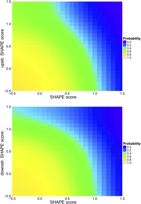
Selective 2'-hydroxyl acylation analyzed by primer extension (SHAPE) is a facile technique for quantitative analysis of RNA secondary structure. In general, low SHAPE signal values indicate Watson-Crick base-pairing, and high values indicate positions that are single-stranded within the RNA structure. However, the relationship of SHAPE signals to structural properties such as non-Watson-Crick base-pairing or stacking has thus far not been thoroughly investigated. Here, we present results of SHAPE experiments performed on several RNAs with published three-dimensional structures. This strategy allows us to analyze the results in terms of correlations between chemical reactivities and structural properties of the respective nucleotide, such as different types of base-pairing, stacking, and phosphate-backbone interactions. We find that the RNA SHAPE signal is strongly correlated with cis-Watson-Crick/Watson-Crick base-pairing and is to a remarkable degree not dependent on other structural properties with the exception of stacking. We subsequently generated probabilistic models that estimate the likelihood that a residue with a given SHAPE score participates in base-pairing. We show that several models that take SHAPE scores of adjacent residues into account perform better in predicting base-pairing compared with individual SHAPE scores. This underscores the context sensitivity of SHAPE and provides a framework for an improved interpretation of the response of RNA to chemical modification.


Figures

FIGURE 1.
FIGURE 1.
Examination of RNAs by nondenaturing polyacrylamide gel electrophoresis. They are (1) musD transport element mutant 6 (external control); (2) HDV ribozyme (PDB: 1CX0); (3) signal recognition particle (PDB: 1Z43); (4) HCV IRES (PDB: 1P5O); (5) self-splicing intron (PDB: 1KXK); (6) BWYV pseudoknot (PDB: 437D); (7) tRNA (PDB: 2TRA); and (8) SARS stem–loop II motif (PDB: 1XJR). RNAs were resolved for ∼6 h at 190 V on a 10% nondenaturing polyacrylamide gel in a running buffer containing 5 mM Mg2+. The gel was stained with Fast RNA stain.
FIGURE 2.
FIGURE 2.
Secondary structure of the (A) yeast group II self-splicing intron (PDB 1KXK), (B) BWYV frame shifting pseudoknot (PDB 437D), and (C) SARS stem–loop II motif (PDB 1XJR), color-coded according to SHAPE data. The color-coding is showing the posterior probabilities of cis-Watson-Crick/Watson-Crick base-pairing derived from SHAPE signal values, where blue values correspond to high probabilities (low SHAPE signals) and red values to low probabilities (high SHAPE signals). The probabilistic model was not applied to residues (shown in white) that correspond to the first and last residues for which SHAPE data are available. Residues for which no SHAPE signal values were measured are also shown in white.
FIGURE 3.
FIGURE 3.
Secondary structure of (A) hepatitis delta virus ribozyme (PDB 1CX0), (B) hepatitis C virus IRES domain II (PDB 1P5O), (C) 7S.S SRP RNA (PDB 1Z43), and (D) of yeast tRNAASP (PDB 2TRA) colored according to SHAPE data. Color-coding is as described in the legend of Figure 2.
FIGURE 4.
FIGURE 4.
Density estimation of SHAPE scores that do or do not participate in cis-Watson-Crick/Watson-Crick base-pairing. Narrow distribution indicates base-paired residues; broad distribution, non-base-paired residues.
FIGURE 5.
FIGURE 5.
Bayesian posterior probability of cis-Watson-Crick/Watson-Crick base-pairing as a function of SHAPE score. Indicated in gray is a 95% confidence interval. The method for estimating the posterior probability as well as the confidence interval is described in the Supplemental Material (sections 1 and 2).
FIGURE 6.
FIGURE 6.
Bayesian posterior probability of cis-Watson-Crick/Watson-Crick base-pairing of a residue as a function of its SHAPE score and the SHAPE score of one adjacent residue. (Top) The upstream adjacent residue is chosen; (bottom) the downstream adjacent residue is chosen.

References

    1. Badorrek CS, Weeks KM 2005. RNA flexibility in the dimerization domain of a gamma retrovirus. Nat Chem Biol 1: 104–111 - PubMed
    1. Darty K, Denise A, Ponty Y 2009. VARNA: Interactive drawing and editing of the RNA secondary structure. Bioinformatics 25: 1974–1975 - PMC - PubMed
    1. Das R, Laederach A, Pearlman SM, Herschlag D, Altman RB 2005. SAFA: semi-automated footprinting analysis software for high-throughput quantification of nucleic acid footprinting experiments. RNA 11: 344–354 - PMC - PubMed
    1. Deigan KE, Li TW, Mathews DH, Weeks KM 2009. Accurate SHAPE-directed RNA structure determination. Proc Natl Acad Sci 106: 97–102 - PMC - PubMed
    1. Geissmann T, Marzi S, Romby P 2009. The role of mRNA structure in translational control in bacteria. RNA Biol 6: 153–160 - PubMed

Publication types

LinkOut - more resources