. 2011 Aug;138(16):3421-30.
doi: 10.1242/dev.068601. Epub 2011 Jul 13.

The regenerative capacity of zebrafish reverses cardiac failure caused by genetic cardiomyocyte depletion

Affiliations

The regenerative capacity of zebrafish reverses cardiac failure caused by genetic cardiomyocyte depletion

Jinhu Wang et al. Development. 2011 Aug.

Abstract

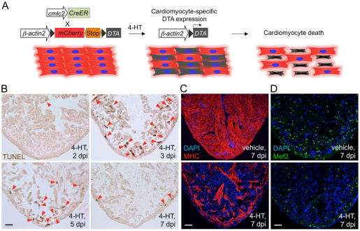
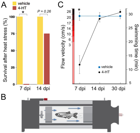
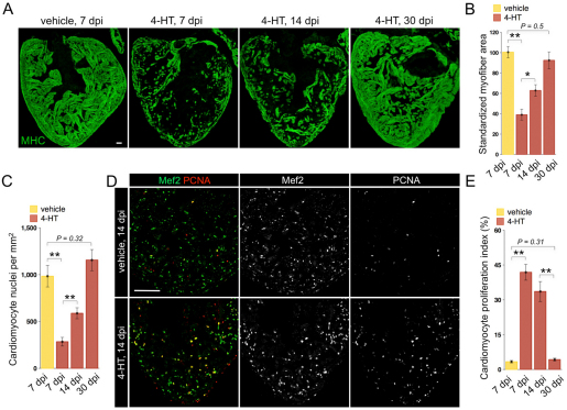
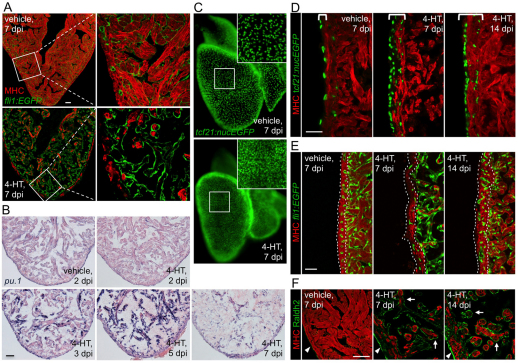
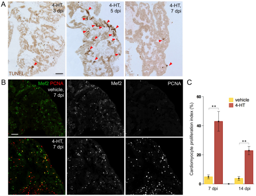
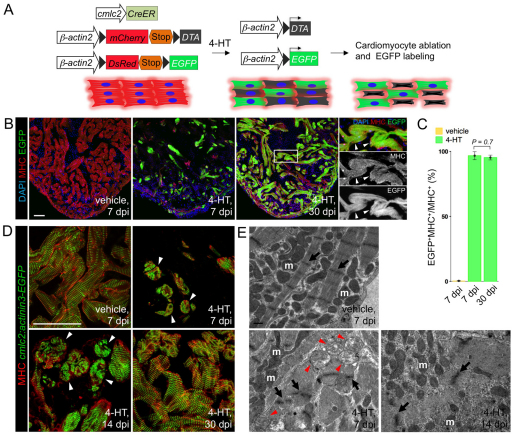
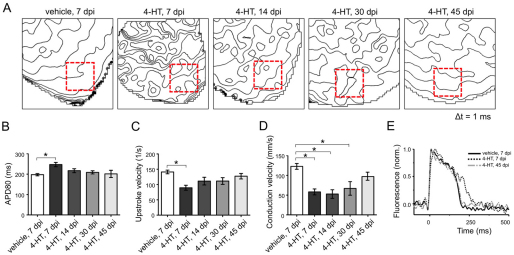
Natural models of heart regeneration in lower vertebrates such as zebrafish are based on invasive surgeries causing mechanical injuries that are limited in size. Here, we created a genetic cell ablation model in zebrafish that facilitates inducible destruction of a high percentage of cardiomyocytes. Cell-specific depletion of over 60% of the ventricular myocardium triggered signs of cardiac failure that were not observed after partial ventricular resection, including reduced animal exercise tolerance and sudden death in the setting of stressors. Massive myocardial loss activated robust cellular and molecular responses by endocardial, immune, epicardial and vascular cells. Destroyed cardiomyocytes fully regenerated within several days, restoring cardiac anatomy, physiology and performance. Regenerated muscle originated from spared cardiomyocytes that acquired ultrastructural and electrophysiological characteristics of de-differentiation and underwent vigorous proliferation. Our study indicates that genetic depletion of cardiomyocytes, even at levels so extreme as to elicit signs of cardiac failure, can be reversed by natural regenerative capacity in lower vertebrates such as zebrafish.


Figures

Fig. 1.
Fig. 1.
Genetic ablation of adult zebrafish cardiomyocytes. (A) Schematic representation of transgenes used for zebrafish cardiomyocyte ablation. DTA is specifically expressed in cardiomyocytes upon 4-HT treatment. (B) TUNEL staining of ventricular muscle from transgenic animals injected with vehicle or 4-HT (0.5 mg/ml). Arrowheads indicate TUNEL-positive cardiac myocytes. (C) Myosin heavy chain (MHC) staining of ventricular sections after vehicle or 4-HT injection, shown at 7 days post-injection (dpi). (D) Mef2 staining to indicate cardiomyocyte nuclei, showing major cellular losses in 4-HT-injected animals. Scale bars: 50 μm.
Fig. 2.
Fig. 2.
Stress response and swimming performance after cardiomyocyte ablation. (A) Survival of Z-CAT after a strong heat shock is given 7 and 14 days after vehicle or 4-HT injection. For each group, 9-13 zebrafish were assessed. Fisher Irwin exact test, *P<0.05. (B) Cartoon representing assay for swimming performance, in which adult zebrafish must swim to maintain position in a controlled current. (C) Maximum swimming speed of Z-CAT fish injected with vehicle or 4-HT at 7, 14 and 30 days post-injection (dpi). At each time point, 7-15 animals were assessed. Several vehicle-treated animals maintained swimming orientation throughout the 32-minute test periods; thus, 7 dpi data under-represent differences between the groups. *P<0.05, Student's t-test. Mean±s.e.m.
Fig. 3.
Fig. 3.
Rapid regeneration of ventricular cardiomyocytes after ablation-induced injuries. (A) Myosin heavy chain (MHC) staining of ventricular sections from Z-CAT fish injected with vehicle or 4-HT, at 7, 14 and 30 days post-injection (dpi). For each group, 5-7 animals were assessed. (B) Quantification of MHC+ myofiber area from experiments in A. (C) Quantification of Mef2+ cardiomyocyte nuclei from experiments in A. (D) Ventricular cardiomyocyte proliferation at 14 dpi assessed by Mef2 and PCNA staining. 4-HT-injected animals display widespread PCNA+ cardiomyocytes. (E) Quantification of ventricular cardiomyocyte proliferation in Z-CAT animals injected with vehicle or 4-HT, at 7, 14 and 30 dpi. For each group, seven to nine animals were assessed. *P<0.05, **P<0.005, Student's t-test. Mean±s.e.m. Scale bars: 50 μm.
Fig. 4.
Fig. 4.
Effects of cardiomyocyte ablation on non-muscle cells. (A) Visualization of endocardial cells (green) and muscle (red) in sections from vehicle-or 4-HT-injected Z-CAT fish. The endocardial layer appears intact one week after massive cardiomyocyte loss. Single confocal slices are shown. (B) In situ hybridization for pu.1-expressing cells, which are prominent by 3-5 dpi. (C) Whole-mount visualization of epicardial nuclei in vehicle- or 4-HT-injected Z-CAT fish at 7 dpi, indicating higher epicardial cell density after myocardial injury. Inset shows enlarged view of boxed area. (D) Histological visualization of epicardial cells after myocardial ablation. Epicardial cells proliferate by 7 dpi and are incorporated into the regenerating myocardial wall by 14 dpi. Brackets indicate area containing tcf21-expressing epicardial cells. (E) Visualization of coronary vascular endothelial cells (green) in sections of the ventricular wall (outlined by dotted lines). Coronary vasculature is reduced in 4-HT-injected Z-CAT animals by 7 dpi, but restored by 14 dpi. (F) Sections from Z-CAT ventricles stained for the retinoic acid (RA)-synthesizing enzyme Raldh2. RA synthesis appears low in vehicle-injected tissue, but is strongly induced in epicardial (arrowheads) and endocardial (arrows) structures after cardiomyocyte ablation. Single confocal slices are shown. Scale bars: 50 μm.
Fig. 5.
Fig. 5.
Atrial cardiomyocyte ablation and regeneration. (A) TUNEL staining of atrial sections from Z-CAT fish injected with 4-HT. Arrowheads indicate TUNEL-positive muscle. (B) Cardiomyocyte proliferation at 7 days post-injection (dpi) in atrial sections from Z-CAT animals injected with vehicle or 4-HT, assessed by Mef2 and PCNA staining. 4-HT-injected animals display widespread atrial PCNA+ cardiomyocytes. Scale bars: 50 μm. (C) Quantification of atrial cardiomyocyte proliferation in Z-CAT fish injected with vehicle or 4-HT, at 7 and 14 dpi. For each group, six to eight animals were assessed. Mean±s.e.m. **P<0.005, Student's t-test.
Fig. 6.
Fig. 6.
Lineage-tracing and ultrastructural analysis of regenerating muscle. (A) Schematic representation of transgenes used for myocyte ablation and lineage tracing. (B) Triple transgenic (Z-CAT; bactin2:loxp-DsRed-STOP-loxp-EGFP) zebrafish were injected once with vehicle (left) or 4-HT. In 4-HT-injected animals, EGFP labeled the majority of spared cardiomyocytes by 7 dpi (middle) and the majority of regenerated cardiomyocytes by 30 days post-injection (dpi; right). Arrowheads indicate examples of MHC+ tissue expressing EGFP. (C) Quantification of EGFP+ myocardium as a percentage of MHC+ ventricular tissue. At 7 dpi, 96% of myocardium was EGFP-labeled, and 95% at 30 dpi. For each group, 4-10 animals were assessed. Insets show enlarged views of the boxed area. (D) Assessment of sarcomere structure in Z-CAT fish crossed to a transgenic strain that labels Z-lines (green). Cardiomyocytes of control animals show well-organized sarcomeres with clear Z-lines. By contrast, many myocytes of 4-HT-injected animals display disorganized sarcomeres and loss of Z-lines (arrowheads) by 7 dpi and 14 dpi. By 30 dpi, sarcomeric structure and Z-lines are typically restored. Scale bars: 50 μm. (E) Transmission electron micrographs of ventricular myocytes from vehicle- or 4-HT-injected Z-CAT animals. Control myocytes have prominent sarcomeric structure and normal mitochondria (m), whereas 7 and 14 dpi myocytes appear less organized with fewer Z-lines (arrows) and swollen mitochondria (arrowheads). Scale bar: 0.5 μm.
Fig. 7.
Fig. 7.
Electrical properties of ventricular cardiomyocytes after widespread ablation. (A) Isochronal maps of ventricular apices from vehicle- or 4-HT-injected Z-CAT fish at indicated time points display the position of the action potential wavefront at 1 ms intervals (Δt=1 ms). Squares show regions of interest (ROIs; 156×156 μm) for epicardial surface conduction velocity estimation shown in D. (B,C) Action potential duration estimated from 20% depolarization to 80% repolarization (APD80) (B), and derived maximum upstroke velocities (C). Mean±s.e.m. *P<0.05, at 7 dpi and non-significant (NS) at 14, 30 and 45 dpi; one-way ANOVA with Tukey's HSD for comparisons with ventricles from vehicle-injected fish. (D) Mean estimated surface myocardium conduction velocities from ROIs in A. Mean±s.e.m. *P<0.05 at 7, 14 and 30 dpi and NS at 45 dpi; one-way ANOVA with Tukey's HSD for comparisons with ventricles from vehicle-injected fish. (E) Representative traces of surface action potentials, indicating a slowing of the maximum depolarization rate and an increase in action potential duration at 7 dpi, and their recovery at 45 dpi.

References

    1. Akazawa H., Komazaki S., Shimomura H., Terasaki F., Zou Y., Takano H., Nagai T., Komuro I. (2004). Diphtheria toxin-induced autophagic cardiomyocyte death plays a pathogenic role in mouse model of heart failure. J. Biol. Chem. 279, 41095-41103 - PubMed
    1. Beltrami A. P., Barlucchi L., Torella D., Baker M., Limana F., Chimenti S., Kasahara H., Rota M., Musso E., Urbanek K., et al. (2003). Adult cardiac stem cells are multipotent and support myocardial regeneration. Cell 114, 763-776 - PubMed
    1. Beltrami C. A., Finato N., Rocco M., Feruglio G. A., Puricelli C., Cigola E., Quaini F., Sonnenblick E. H., Olivetti G., Anversa P. (1994). Structural basis of end-stage failure in ischemic cardiomyopathy in humans. Circulation 89, 151-163 - PubMed
    1. Bersell K., Arab S., Haring B., Kuhn B. (2009). Neuregulin1/ErbB4 signaling induces cardiomyocyte proliferation and repair of heart injury. Cell 138, 257-270 - PubMed
    1. Braunwald E., Bonow R. O. (2010). Braunwald's Heart Disease: A Textbook of Cardiovascular Medicine. Philadelphia: Saunders;

Publication types