. 2011 Aug;138(16):3367-76.
doi: 10.1242/dev.065797. Epub 2011 Jul 13.

Live imaging of the Drosophila spermatogonial stem cell niche reveals novel mechanisms regulating germline stem cell output

Affiliations

Live imaging of the Drosophila spermatogonial stem cell niche reveals novel mechanisms regulating germline stem cell output

X Rebecca Sheng et al. Development. 2011 Aug.

Abstract

Adult stem cells modulate their output by varying between symmetric and asymmetric divisions, but have rarely been observed in living intact tissues. Germline stem cells (GSCs) in the Drosophila testis are anchored to somatic hub cells and were thought to exclusively undergo oriented asymmetric divisions, producing one stem cell that remains hub-anchored and one daughter cell displaced out of the stem cell-maintaining micro-environment (niche). We developed extended live imaging of the Drosophila testis niche, allowing us to track individual germline cells. Surprisingly, new wild-type GSCs are generated in the niche during steady-state tissue maintenance by a previously undetected event we term 'symmetric renewal', where interconnected GSC-daughter cell pairs swivel such that both cells contact the hub. We also captured GSCs undergoing direct differentiation by detaching from the hub. Following starvation-induced GSC loss, GSC numbers are restored by symmetric renewals. Furthermore, upon more severe (genetically induced) GSC loss, both symmetric renewal and de-differentiation (where interconnected spermatogonia fragment into pairs while moving towards then establishing contact with the hub) occur simultaneously to replenish the GSC pool. Thus, stereotypically oriented stem cell divisions are not always correlated with an asymmetric outcome in cell fate, and changes in stem cell output are governed by altered signals in response to tissue requirements.


Figures

Fig. 1.
Fig. 1.
Germ cells appear healthy and divide in live imaged testes. (A) Schematic of the testis apex depicting hub cells (blue), GSCs (green), mitotic spindle (pink), centrosomes (white), spermatogonia and gonialblasts (yellow), cyst stem cells (dark grey) and cyst cells (light gray). (B) Graph comparing the relative frequencies of dividing germ cells of a particular stage in fixed and live imaged testes using the Chi-squared test. (C-F) Frames and corresponding schematics showing GSC division in a time-lapse imaged testis expressing GMA in GSCs and spermatogonia (genotype: Hs-bam; nanos-Gal4; UAS-GMA/+). (C) Before division occurs, the GSC (arrowhead, green in schematic) has GMA enriched at the stem cell-hub interface. (D) During mitosis, GMA becomes highly enriched at the cell cortex, and later marks the cleavage furrow during incomplete cytokinesis. (E,F) A daughter gonialblast (arrow, yellow) is formed (E) and remains displaced from the hub until the end of the movie (F). Images are maximum intensity projections of three 1.5 μm optical sections. Illustrations depicting the cells of interest are below their respective panels. The hub is outlined. Scale bar: 10 μm.
Fig. 2.
Fig. 2.
GSC division can result in the production of a GSC and GB (asymmetric division), two GSCs (symmetric renewal) or the loss of a GSC (symmetric differentiation). (A-L) Time-lapse images taken from a movie of P{nos::EGFP-moe::nos 3UTR} testis. (A-D) Example of a GSC division that results in a daughter cell (arrow) remaining displaced away from the hub until the end of the movie. Arrowhead indicates daughter cell. (E-H) GSC-daughter pairs (arrowhead, arrow) can swivel such that the initially displaced daughter contacts and is maintained at the hub. (I-L) GSC-daughter pairs (arrowhead, arrow) can detach from the hub and remain morphologically indistinguishable from two-cell spermatogonial clusters for the remainder of the movie. Illustrations representing the cells of interest are below their respective panels with colors corresponding to their function as suggested by their anatomical location: green, GSC; yellow, daughter cell. Asterisks indicate the hub. Scale bar: 10 μm.
Fig. 3.
Fig. 3.
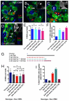
GSCs undergoing symmetric renewal are detected in fixed tissues. (A-C) Testes of genotype P{nos::EGFP-moe::nos 3UTR} were incubated in media containing EdU for 20 minutes, fixed and then visualized for EdU (red) and immunostained for GMA (green), Armadillo and 1B1 (blue). (A) Two labeled GSCs (solid arrowheads) both attached to labeled daughter cells (arrows). (B) A labeled GSC-daughter pair with both cells attached to the hub (solid arrowheads) and fusome (open arrowhead; inset shows the blue channel) between the cells. No single daughter cells were nearby, suggesting that abscission had not occurred prior to fixation. (C) An EdU-labeled GSC-daughter pair (arrows) displaced from the hub with a long GMA-rich protrusion still attached at the hub (open arrowhead; inset shows the green channel). (D) A GSC-daughter pair (solid arrowheads) with both cells at the hub in Cnn HK21/mfs3 trans-heterozygotes immunostained with the germ cell marker Vasa (green) instead of GFP. Scale bar: 10 μm. (E) The number of GSCs is not significantly different between heterozygous controls and trans-heterozygous or homozygous centrosomin mutants (n=number of testes). (F) The percentage of EdU labeled GSCs with daughters adjacent to the hub was significantly increased in centrosomin mutants, including the heterozygous HK21 allele (n=number of GSCs). (G) Schematic of starvation and feeding protocol performed on P{nos::EGFP-moe::nos 3UTR} flies; broken lines represent time spent on 10% sucrose food and unbroken lines represent time spent on standard molasses food. (H) Starvation causes a modest but significant decrease in the number of GSCs compared with age-matched controls, followed by recovery upon feeding (n=number of testes). (I) The percentage of GSC-daughter pairs with both cells at the hub increases after starvation and after 1 day of feeding, but returns to baseline after 5 days of feeding (n=number of GSCs). Error bars represent s.d. Statistical analysis of GSC number performed using ANOVA with Dunn's and Tukey's multiple comparison tests (*P<0.05). Statistical analysis comparing percentages performed using Chi-squared test.
Fig. 4.
Fig. 4.
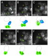
Live imaging shows de-differentiating spermatogonia moving to make contact with the niche and also undergoing symmetric renewal. (A-F) Time-lapse images taken from movie of Hs-bam; Nanos-Gal4; UAS-GMA testes depleted of GSCs and allowed to recover for 36 hours before imaging. Numbered cells and hub (dotted circle) are depicted in illustrations. (A) Two pairs of germ cells (1,2) and (3,4), with GMA enrichment between the syncytial cells, are initially not in contact with the hub. (B) Pair (1,2) moves first such that both cells make large hub contacts, (C) followed by pair (3,4). Cells 1 and 3 are contacting the hub, but out of the focal plane. (D) Cell 4, which had attached to the hub, divides to produce daughter 4a. (E) 4a initially has a very little hub contact; (F) this gradually increases. This is an example of a division where the daughter cell is not displaced from the hub. Images are maximum intensity projections of three 1.5 μm optical sections. The hub is indicated by an asterisk. Scale bar: 10 μm.
Fig. 5.
Fig. 5.
The outcomes of GSC division are significantly altered between steady-state and regeneration. (A) Graph showing the 119 events observed during steady-state and 102 events during regeneration categorized as asymmetric division, symmetric renewal, loss or reversion. The total number of events is derived from the number of observed GSC mitoses (regardless of division orientation) plus the number of cases of spermatogonial reversion, symmetric renewal and symmetric differentiation that occurred without an observed GSC division. GSCs that undergo mitosis and produce daughter cells that remained displaced away from the hub until the end of imaging were scored as asymmetric division. GSC-daughter pairs that swivel such that the daughter cell makes and maintains contact with the hub throughout the remaining imaging period (this includes cases where a GSC divided parallel or 45° to the hub) were scored as symmetric renewals. GSC-daughter pairs detaching from the hub and remaining detached throughout the subsequent imaging period were scored as symmetric differentiation (loss). Gonialblasts, two-, four-, eight- or 16-cell spermatogonial clusters moving from not being in direct contact to being attached to the hub were scored as reversions. Chi-squared analysis reveals the proportions of the events to be significantly different. (B) Schematic showing relative frequency of events during steady-state and regeneration.

References

    1. Barroca V., Lassalle B., Coureuil M., Louis J. P., Le Page F., Testart J., Allemand I., Riou L., Fouchet P. (2009). Mouse differentiating spermatogonia can generate germinal stem cells in vivo. Nat. Cell Biol. 11, 190-196 - PubMed
    1. Bloor J. W., Kiehart D. P. (2001). zipper Nonmuscle myosin-II functions downstream of PS2 integrin in Drosophila myogenesis and is necessary for myofibril formation. Dev. Biol. 239, 215-228 - PubMed
    1. Boyle M., Wong C., Rocha M., Jones D. L. (2007). Decline in self-renewal factors contributes to aging of the stem cell niche in the Drosophila testis. Cell Stem Cell 1, 470-478 - PubMed
    1. Brawley C., Matunis E. (2004). Regeneration of male germline stem cells by spermatogonial dedifferentiation in vivo. Science 304, 1331-1334 - PubMed
    1. Carpenter A. T. (1981). EM autoradiographic evidence that DNA synthesis occurs at recombination nodules during meiosis in Drosophila melanogaster females. Chromosoma 83, 59-80 - PubMed

Publication types