

Varying effects of different β-glucans on the maturation of porcine monocyte-derived dendritic cells
- PMID: 21752950
- PMCID: PMC3165223
- DOI: 10.1128/CVI.00080-11
Varying effects of different β-glucans on the maturation of porcine monocyte-derived dendritic cells
Abstract
β-Glucans are well known for their immunomodulatory capacities in humans and mice. For this reason, together with the European ban on growth-promoting antibiotics, β-glucans are intensively used in pig feed. However, as shown in the present study, there is much variation in the stimulatory capacities of β-glucans from different sources. Since dendritic cells (DCs) are the first cells that are encountered after an antigen is taken up by the intestinal epithelial cell barrier, we decided to investigate the effect of two concentrations (5 and 10 μg/ml) of five commercial β-glucan preparations, differing in structure and source, on porcine monocyte-derived dendritic cells (MoDCs). Although all β-glucans gave rise to a significant reduction of the phagocytic activity of DCs, only Macrogard induced a significant phenotypic maturation. In addition to Macrogard, zymosan, another β-glucan derived from Saccharomyces cerevisiae, and curdlan also significantly improved the T-cell-stimulatory capacity of MoDCs. Most interesting, however, is the cytokine secretion profile of curdlan-stimulated MoDCs, since only curdlan induced significant higher expression levels of interleukin-1β (IL-1β), IL-6, IL-10, and IL-12/IL-23p40. Since the cytokine profile of DCs influences the outcome of the ensuing immune response and thus may prove valuable in intestinal immunity, a careful choice is necessary when β-glucans are used as dietary supplement.
Figures
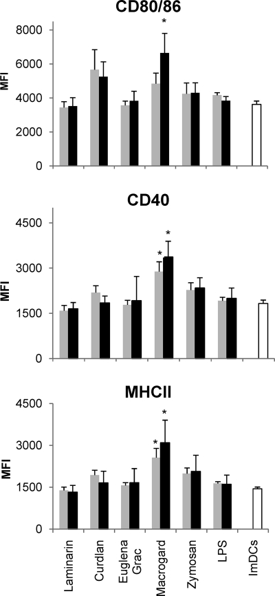
 ) or 10 μg (▪) of β-glucan/ml or LPS (1 and 10 μg/ml). The expression of the maturation markers was assayed by flow cytometry. The data are shown as the means ± the standard errors of the mean (SEM) for four pigs. Asterisks (*) indicate a significant difference between β-glucan-stimulated MoDCs and immature MoDCs (ImDCs) (P < 0.01).
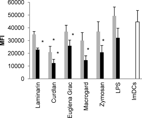 ) or 10 μg (▪) of β-glucan/ml or LPS (1 and 10 μg/ml), and the uptake of ova-dQ was assayed by flow cytometry. Mean fluorescence intensity (MFI) values were calculated by subtracting the MFI values obtained at 4°C from those obtained at 37°C. The data are shown as the means ± the SEM for four pigs. Asterisks (*) indicate a significant difference between β-glucan-stimulated MoDCs and immature MoDCs (ImDCs) (for all β-glucans P < 0.01 except for the β-glucan from Euglena gracilis).
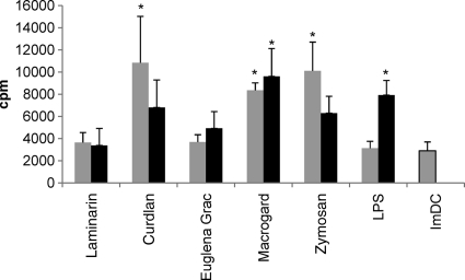 ) or 10 μg (▪) of different β-glucans/ml or LPS (1 or 10 μg/ml) and then added to CD172a-depleted PBMC (lymphocytes). After 5 days, the cultures were pulsed with 1 μCi of [3H]methyl-thymidine. T-cell proliferation was measured after an additional coculture of 18 h. The data are shown as means ± the SEM for four pigs. Asterisks (*) indicate a significant difference between β-glucan-stimulated conditions and the untreated condition (ImDCs) (P < 0.01 except for LPS [10 μg/ml]; P < 0.05). The proliferative responses of the CD172a− lymphocytes (i.e., no MoDCs were added) was less than 300 cpm.
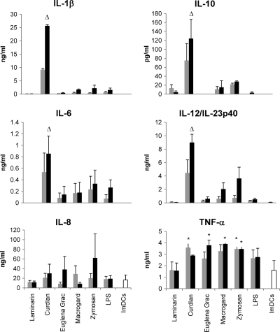 ) and 10 μg (▪) of different β-glucans/ml or LPS (1 or 10 μg/ml). After 24 h, the culture supernatant was harvested, and the IL-1β, IL-6, IL-8, IL-10, IL-12/IL-23p40, and TNF-α cytokine concentrations were measured using commercially available ELISA kits. The data are shown as means ± the SEM for four pigs. Asterisks (*) indicate a significant difference (P < 0.05) between β-glucan-stimulated MoDCs and immature MoDCs, while a triangle (▵) indicates a significant difference (P < 0.01) between MoDCs stimulated with 10 μg of curdlan/ml and all of the other β-glucan-stimulated (10 μg/ml) MoDCs or immature MoDCs.
References
-
- Backer R., van Leeuwen F., Kraal G., den Haan J. M. 2008. CD8− dendritic cells preferentially cross-present Saccharomyces cerevisiae antigens. Eur. J. Immunol. 38:370–380 - PubMed
-
- Bimczok D., et al. 2007. Cholera toxin promotes the generation of semi-mature porcine monocyte-derived dendritic cells that are unable to stimulate T cells. Vet. Res. 38:597–612 - PubMed
Publication types
MeSH terms
Substances
LinkOut - more resources
Full Text Sources
