. 2011 Sep;60(9):2331-40.
doi: 10.2337/db11-0159. Epub 2011 Jul 13.

Immunosuppressive effects of streptozotocin-induced diabetes result in absolute lymphopenia and a relative increase of T regulatory cells

Affiliations

Immunosuppressive effects of streptozotocin-induced diabetes result in absolute lymphopenia and a relative increase of T regulatory cells

Yannick D Muller et al. Diabetes. 2011 Sep.

Abstract

Objective: Streptozotocin (STZ) is the most widely used diabetogenic agent in animal models of islet transplantation. However, the immunomodifying effects of STZ and the ensuing hyperglycemia on lymphocyte subsets, particularly on T regulatory cells (Tregs), remain poorly understood.

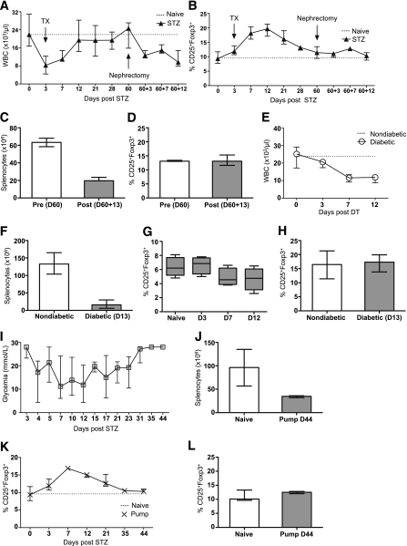
Research design and methods: This study evaluated how STZ-induced diabetes affects adaptive immunity and the consequences thereof on allograft rejection in murine models of islet and skin transplantation. The respective toxicity of STZ and hyperglycemia on lymphocyte subsets was tested in vitro. The effect of hyperglycemia was assessed independently of STZ in vivo by the removal of transplanted syngeneic islets, using an insulin pump, and with rat insulin promoter diphtheria toxin receptor transgenic mice.

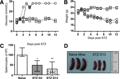
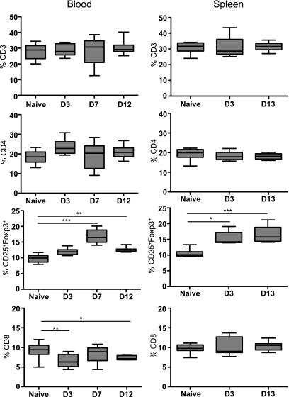
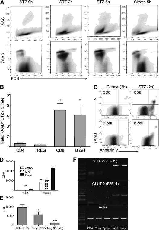
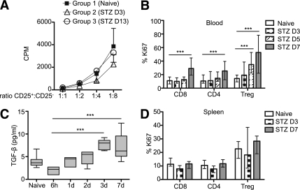
Results: Early lymphopenia in both blood and spleen was demonstrated after STZ administration. Direct toxicity of STZ on lymphocytes, particularly on CD8(+) cells and B cells, was shown in vitro. Hyperglycemia also correlated with blood and spleen lymphopenia in vivo but was not lymphotoxic in vitro. Independently of hyperglycemia, STZ led to a relative increase of Tregs in vivo, with the latter retaining their suppressive capacity in vitro. The higher frequency of Tregs was associated with Treg proliferation in the blood, but not in the spleen, and higher blood levels of transforming growth factor-β. Finally, STZ administration delayed islet and skin allograft rejection compared with naive mice.

Conclusions: These data highlight the direct and indirect immunosuppressive effects of STZ and acute hyperglycemia, respectively. Thus, these results have important implications for the future development of tolerance-based protocols and their translation from the laboratory to the clinic.


Figures

FIG. 1.
FIG. 1.
Diabetes induction with STZ diminishes the absolute numbers of splenocytes. A: Blood glucose levels (mmol/L). ○, STZ-induced diabetic mice; □, naive mice. B: Weight (g) of C57BL/6 mice after STZ administration. ○, STZ-induced diabetic mice; □, naive mice. C: Absolute numbers of splenocytes 3 (D3) and 13 (D13) days after STZ compared with naive mice (n = 5). The median and the range are shown and were analyzed with a nonparametric Kruskal-Wallis test. D: Three spleens of STZ-induced diabetic mice at day 13 compared with naive mice are shown. A P value inferior to 0.05 was considered statistically significant (*P < 0.05, **P < 0.01).
FIG. 2.
FIG. 2.
Flow cytometry analysis of T cells in STZ-induced diabetic mice. CD3+, CD4+, CD25+Foxp3+ (gated in the CD4+ population), and CD8+ cell percentages were analyzed in the blood at day 3 (D3), 7 (D7), and 12 (D12) and in the spleen at days 3 (D3) and 13 (D13). Blood from 15 naive and 8 STZ-induced diabetic mice was analyzed. Spleens of eight naive and five STZ-induced diabetic mice were analyzed. Median and the range are shown. A nonparametric Kruskal-Wallis test was performed. *P < 0.05, **P < 0.01, ***P < 0.001.
FIG. 3.
FIG. 3.
The effects of STZ and acute hyperglycemia. The respective effects of STZ and hyperglycemia were analyzed in three different models. First (A–D), C57BL/6 mice were transplanted 3 days after STZ administration with 400 islets equivalent under the left kidney capsule. Sixty days after Tx, a nephrectomy (graftectomy) was performed to induce diabetes without STZ. Absolute numbers of white blood cells (A) and percentage of CD25+Foxp3+ Tregs gated in the CD4+ population (B) were assessed at different time points in the blood (days 0, 3, 7, 12, 21, 28, and 60 after STZ administration and 60 + 3, 60 + 7, and 60 + 12 after nephrectomy). Absolute numbers of splenocytes (C) and percentage of CD25+Foxp3+ Tregs (D) were assessed in the spleen before nephrectomy (day 60 [D60]) and after nephrectomy (day 60 + 13 [D60+13]). Second (E–H), RIP-DTR transgenic mice were made diabetic by administration of DT. Absolute numbers of leukocytes were assessed in the blood at days 3, 7, and 12 (E) and in the spleen at day 13 (D13) after DT administration (F). Percentage of Tregs in RIP-DTR transgenic mice was assessed at the same time points in the blood (G) and in the spleen (H). Third (I–L), 3 days after STZ administration, an insulin pump was implanted subcutaneously to control the glycemia for ∼20 days. Thereafter, the insulin pump was ineffective, and the mice became diabetic again (I). Absolute numbers of splenocytes were assessed at day 44 (D44) after STZ (J). Treg percentage was calculated in the blood at days 3, 7, 12, 21, 35, and 44 (K) and in the spleen at day 44 (D44) (L). The median and the range are shown.
FIG. 4.
FIG. 4.
Effect of STZ on leukocytes in vitro. A: Splenocytes were harvested from Foxp3 GFP knock-in mice and cultured for 1 h in 4.4 mmol/L STZ or control citrate-conditioned medium (10% FCS). 7AAD+ cells were analyzed by flow cytometry 0, 2, and 5 h after STZ exposition. One representative experiment out of four is shown. B: Percentage of 7AAD+ cells in CD4+, Foxp3+, CD8+, and B cells were further analyzed 2 h after STZ or citrate exposition to determine which cell subtypes were more sensitive to STZ. The results were calculated as followed: % of 7AAD+ cells (STZ condition) divided by percent of 7AAD+ cells (citrate condition). The mean and SD of four separate experiments pooled together are shown and analyzed by one-way ANOVA. C: CD8+ and B220+ cells were analyzed for Annexin V expression 2 h after STZ or citrate exposition. One representative experiment out of four is shown. D: After exposition to STZ or citrate-conditioned medium, splenocytes were stimulated with concanavalin A, lipopolysaccharide, or anti-CD3e for 3 days, and their proliferation rate was assessed by [3H]thymidine incorporation. The mean and SD of one representative experiment (performed in triplicate) out of three is shown and analyzed by one-way ANOVA. E: Purified CD4+CD25+ cells were cultured for 1 h in 4.4 mmol/L STZ or citrate-conditioned medium (10% FCS). Thereafter, an in vitro suppression assay was performed. CD4+CD25+ T cells from naive or STZ-induced diabetic mice (day 3 [D3] or 13 [D13]) were added at different CD4+CD25+:CD4+CD25 ratios (1:1, 1:2, 1:4, 1:8). The mean and SD of one representative experiment (performed in triplicate) out of three is shown and analyzed by one-way ANOVA. F: GLUT2 mRNA was analyzed in splenocytes, purified CD4+ cells, and Tregs. Purified islets and liver tissue were used as the control. Two separate experiments were performed. *P < 0.05, ***P < 0.001.
FIG. 5.
FIG. 5.
Treg suppression in vitro and proliferation in vivo. A: CD4+CD25+ cells were purified from naive (group 1) and STZ-induced diabetic mice at day 3 (group 2) (D3) and day 13 (group 3) (D13). The results of an in vitro suppression assay, as described in research design and methods, are shown as mean and SDs. One representative experiment out of three is shown. B: Ki67 expression was analyzed in the blood in CD4+ (total population), CD8+, and Foxp3+ T cells at day 3 (D3), 5 (D5), and 7 (D7) after STZ administration (n = 8–20 per group/time point). C: Plasma TGF-β concentrations were assessed at 6 h and 1, 2, 3, and 7 days (1d, 2d, 3d, 7d) after STZ administration (n = 6). D: Ki67 expression was analyzed in the spleen at days 3 and 7 after STZ administration (n = 6). The median and the range are shown in B, C, and D and were analyzed with the Kruskal-Wallis test. ***P < 0.001.
FIG. 6.
FIG. 6.
Islet and skin allografts in STZ-treated mice. C57BL/6 STZ-induced diabetic (A–C) and naive (D–F) mice were transplanted with BALB/c islets. Islet grafts were analyzed by histology 10 days after Tx. Hematoxylin and eosin (A and D), insulin immunofluorescence (Alexa Fluor 488–conjugated) (B and E), and Foxp3 immunohistochemistry (streptavidin/HRP, black arrows) (C and F) were performed. Magnification is ×200. A–F: Survival curve of BL/6xDBA/2 F1 (H2bxH2d) skin grafts in naive and STZ-induced diabetic mice treated with an implanted insulin pump with or without MR1 treatment. G: Graft survival between groups was compared using the log-rank test. H: Representative skin graft at day 14 after Tx in STZ compared with the naive group. (A high-quality digital representation of this figure is available in the online issue.)

Similar articles

Cited by

References

    1. Lenzen S. The mechanisms of alloxan- and streptozotocin-induced diabetes. Diabetologia 2008;51:216–226 - PubMed
    1. Gaulton GN, Schwartz JL, Eardley DD. Assessment of the diabetogenic drugs alloxan and streptozotocin as models for the study of immune defects in diabetic mice. Diabetologia 1985;28:769–775 - PubMed
    1. Koulmanda M, Qipo A, Auchincloss HJ, Jr, Smith RN. Effects of streptozotocin on autoimmune diabetes in NOD mice. Clin Exp Immunol 2003;134:210–216 - PMC - PubMed
    1. Nichols WK, Spellman JB, Vann LL, Daynes RA. Immune responses of diabetic animals: direct immunosuppressant effects of streptozotocin in mice. Diabetologia 1979;16:51–57 - PubMed
    1. Takayama Y, Ichikawa T, Maki T. Effect of STZ administration on islet isograft and allograft survival in NOD mice. Diabetes 1993;42:324–329 - PubMed

Publication types