. 2011 Jul 13;31(28):10119-27.
doi: 10.1523/JNEUROSCI.1299-11.2011.

Restriction of transient receptor potential vanilloid-1 to the peptidergic subset of primary afferent neurons follows its developmental downregulation in nonpeptidergic neurons

Affiliations

Restriction of transient receptor potential vanilloid-1 to the peptidergic subset of primary afferent neurons follows its developmental downregulation in nonpeptidergic neurons

Daniel J Cavanaugh et al. J Neurosci. .

Abstract

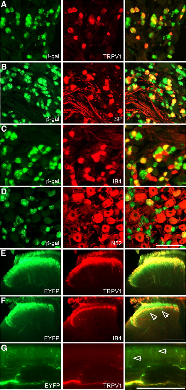
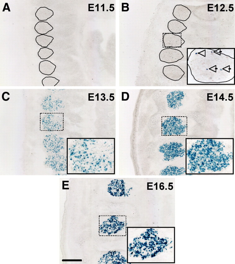
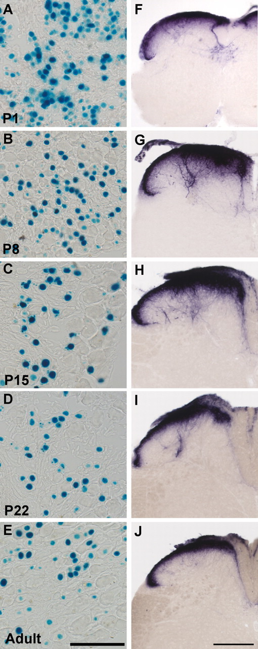
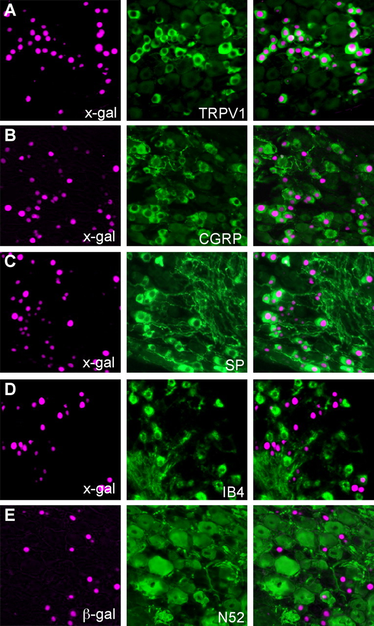
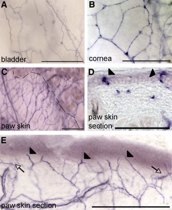
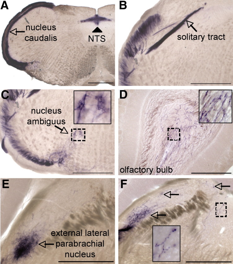
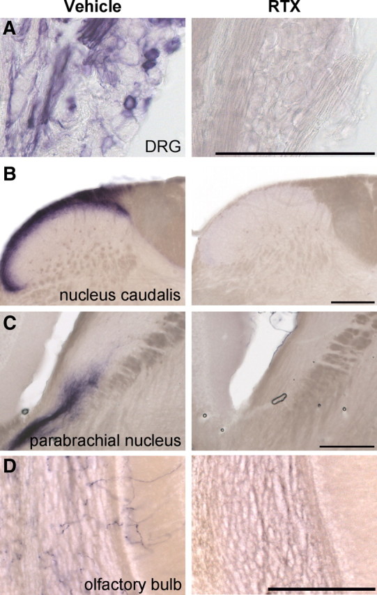
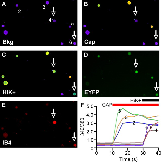
Primary afferent "pain" fibers (nociceptors) are divided into subclasses based on distinct molecular and anatomical features, and these classes mediate noxious modality-specific contributions to behaviors evoked by painful stimuli. Whether the heat and capsaicin receptor transient receptor potential vanilloid-1 (TRPV1) is expressed heterogeneously across several sensory populations, or is selectively expressed by a unique nociceptor subclass, however, is unclear. Here we used two lines of Trpv1 reporter mice to investigate the primary afferent expression of TRPV1, both during development and in the adult. We demonstrate, using Cre-induced lineage tracing, that during development TRPV1 is transiently expressed in a wide range of dorsal root ganglion neurons, and that its expression is gradually refined, such that TRPV1 transcripts become restricted to a specific subset of peptidergic sensory neurons. Finally, the remarkable sensitivity that is characteristic of these reporter mice revealed an innervation of central and peripheral targets by TRPV1+ primary afferents in the adult that is considerably more extensive than has previously been appreciated.


Figures

Figure 1.
Figure 1.
Staining in DRG and spinal cord of TRPV1Cre mice crossed with Cre-dependent reporter lines. A–D, Double labeling of DRG sections from TRPV1Cre/R26R-lacZ mice for lacZ and markers of primary afferent neuron populations. Left panels are stained for lacZ immunoreactivity (green). Middle panels show the same sections stained by immunohistochemistry for the markers listed (red). Right panels show merged images. E, F, Spinal cord sections of TRPV1Cre/R26R-EYFP mice are stained for EYFP (left, green) (E) and TRPV1 or IB4 (middle, red) (F). Merged images are shown on the right. F, Arrowheads show EYFP+ axons extending ventral to the band of IB4 staining. G, Staining in a section of skin from the hindpaw of a TRPV1Cre/R26R-EYFP mouse for EYFP (left, green) and TRPV1 (middle, red). A merged image is shown on the right. Arrowheads show EYFP+/TRPV1−afferent endings. Scale bars: D, G, 100 μm; F, 200 μm.
Figure 2.
Figure 2.
Developmental time course of nlacZ staining in DRG. A–E, nlacZ staining on sections of TRPV1PLAP-nlacZ mouse embryos. A, No nlacZ staining is observed at E11.5 (Dotted lines delineate DRGs). B, By E12.5, a small minority of DRG cells are nlacZ+. Note nlacZ+ cells (arrows). C–E, nlacZ expression increases from E13.5 (C) to E14.5 (D), when the percentage of labeled cells reaches a maximum. D, By E16.5, the overall percentage of nlacZ+ cells has dropped slightly. Insets show magnification of boxed areas. Scale bar: (in E) A–E, 200 μm.
Figure 3.
Figure 3.
Postnatal regulation of TRPV1 in DRG neurons. A–E, nlacZ staining in DRG sections of TRPV1PLAP-nlacZ mice. A, At P1, 59.7 ± 1.7% of DRG neurons were nlacZ+. B, At P8, 45.4 ± 4.4% of DRG neurons were nlacZ+. C–E, By P15, 33.6 ± 4.4% of neurons were nlacZ+ (C), and this percentage stayed constant at P22 (D), and in the adult mouse (E). F–J, PLAP staining of spinal cord sections of TRPV1PLAP-nlacZ mice at P1 (F), P8 (G), P15 (H), (I) P22, and adult (J). Note that staining becomes progressively restricted to the outermost dorsal horn laminae. The percentage of nlacZ+ DRG neurons at each time point is given as mean ± SEM. Scale bars: E, 100 μm; J, 200 μm.
Figure 4.
Figure 4.
Adult expression of TRPV1. A–E, Double labeling of DRG sections from TRPV1PLAP-nlacZ mice for nlacZ and markers of primary afferent neuron populations. A–E, Left panels show nlacZ staining with the X-gal reaction (pseudocolored magenta). Middle panels show the same sections stained by immunohistochemistry for the markers listed. Right panels show merged images.
Figure 5.
Figure 5.
PLAP staining in peripheral tissues targeted by DRG neurons. A, PLAP staining in whole-mount bladder. B, PLAP staining in whole-mount cornea. C, PLAP staining in whole-mount paw skin. Dashed line shows boundary between hairy (right) and glabrous (left) skin. D, In 14 μm sections of paw skin, we occasionally observed PLAP+ fibers extending into the outermost layer of the epidermis (arrowheads). E, PLAP+ afferent fibers were generally observed running parallel to the dermal/epidermal border (arrows) and regularly extended into the innermost epidermal layers (arrowheads). Scale bars: A–C, 200 μm; D, E, 100 μm.
Figure 6.
Figure 6.
PLAP staining in central tissues targeted by DRG neurons. A, PLAP staining in nucleus caudalis (arrow) and the nucleus of the solitary tract (arrowhead) of the medulla. B, Robust PLAP staining in the solitary tract (arrow). C, PLAP staining in the ventral medulla (inset shows magnification of boxed area, near nucleus ambiguus). D, PLAP+ axons in the granule layer of the olfactory bulb (inset shows magnification of boxed area). E, External lateral parabrachial nucleus receives extensive PLAP+ innervation (arrow). F, PLAP+ fibers extend throughout the parabrachial nucleus (arrows). Insets show magnification of boxed areas. Scale bars, 500 μm.
Figure 7.
Figure 7.
Systemic RTX injection elminates PLAP staining in spinal cord and brain. A–D, PLAP staining in DRG (A), nucleus caudalis (B), parabrachial nucleus (C), and olfactory bulb (D) is eliminated following RTX injection. Left panels show PLAP staining in vehicle-injected control mice, and right panels show equivalent areas in RTX-injected mice. Scale bars, 200 μm.
Figure 8.
Figure 8.
Calcium imaging of capsaicin responsiveness in cultured TRPV1Cre/R26R-EYFP DRG neurons. A–C, Cultured DRG neurons from 6-week-old mice were imaged with Fura-2-AM dye at background (A), and following stimulation with 1 or 50 μm capsaicin (B), and high-K+ Ringer's solution (C). D, Endogenous EYFP signal from cells recorded in A–C. E, Cells were then fixed and assayed for IB4 binding. E, 340/380 ratios of cells as numbered in A. Arrows point to EYFP+, IB4-binding cells that were unresponsive to capsaicin. Scale bar, 100 μm.

References

    1. Basbaum AI, Bautista DM, Scherrer G, Julius D. Cellular and molecular mechanisms of pain. Cell. 2009;139:267–284. - PMC - PubMed
    1. Bates BD, Mitchell K, Keller JM, Chan CC, Swaim WD, Yaskovich R, Mannes AJ, Iadarola MJ. Prolonged analgesic response of cornea to topical resiniferatoxin, a potent TRPV1 agonist. Pain. 2010;149:522–528. - PMC - PubMed
    1. Bráz JM, Basbaum AI. Triggering genetically-expressed transneuronal tracers by peripheral axotomy reveals convergent and segregated sensory neuron-spinal cord connectivity. Neuroscience. 2009;163:1220–1232. - PMC - PubMed
    1. Braz JM, Nassar MA, Wood JN, Basbaum AI. Parallel “pain” pathways arise from subpopulations of primary afferent nociceptor. Neuron. 2005;47:787–793. - PubMed
    1. Breese NM, George AC, Pauers LE, Stucky CL. Peripheral inflammation selectively increases TRPV1 function in IB4-positive sensory neurons from adult mouse. Pain. 2005;115:37–49. - PubMed

Publication types