. 2011 Jul 13;31(28):10159-73.
doi: 10.1523/JNEUROSCI.1245-11.2011.

Bcl11b/Ctip2 controls the differentiation of vomeronasal sensory neurons in mice

Affiliations

Bcl11b/Ctip2 controls the differentiation of vomeronasal sensory neurons in mice

Takayuki Enomoto et al. J Neurosci. .

Abstract

The transcription factor Bcl11b/Ctip2 plays critical roles in the development of several systems and organs, including the immune system, CNS, skin, and teeth. Here, we show that Bcl11b/Ctip2 is highly expressed in the developing vomeronasal system in mice and is required for its proper development. Bcl11b/Ctip2 is expressed in postmitotic vomeronasal sensory neurons (VSNs) in the vomeronasal epithelium (VNE) as well as projection neurons and GABAergic interneurons in the accessory olfactory bulb (AOB). In the absence of Bcl11b, these neurons are born in the correct number, but VSNs selectively die by apoptosis. The critical role of Bcl11b in vomeronasal system development is demonstrated by the abnormal phenotypes of Bcl11b-deficient mice: disorganization of layer formation of the AOB, impaired axonal projections of VSNs, a significant reduction in the expression of vomeronasal receptor genes, and defective mature differentiation of VSNs. VSNs can be classified into two major types of neurons, vomeronasal 1 receptor (V1r)/Gα(i2)-positive and vomeronasal 2 receptor (V2r)/Gα(o)-positive VSNs. We found that all Gα(i2)-positive cells coexpressed Gα(o) during embryogenesis. This coexpression is also observed in newly differentiated neurons in the adult VNE. Interestingly, loss of Bcl11b function resulted in an increased number of V1r/Gα(i2)-type VSNs and a decreased number of V2r/Gα(o)-type VSNs, suggesting that Bcl11b regulates the fate choice between these two VSN types. These results indicate that Bcl11b/Ctip2 is an essential regulator of the differentiation and dichotomy of VSNs.


Figures

Figure 1.
Figure 1.
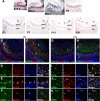
Expression of Bcl11b in the developing mouse vomeronasal epithelium. A, In situ hybridization with RNA probes for Bcl11b in coronal sections of the VNE at E11.5, E12.5, E14.5, E16.5, P0, P3, P7, P14, and P60. The expression of Bcl11b gradually increased during embryogenesis. After birth, the expression of Bcl11b gradually decreased and was restricted to the marginal regions of the VNE in adulthood (arrows). B–M, Bcl11b-expressing cells were characterized using two-color ISH with RNA probes for Bcl11b (red) in combination with marker genes (green) in coronal sections of the VNE at P0 (B–G) and P14 (H–M): Mash1 (neuronal progenitors) (B, H); Ngn1 (neuronal precursors) (C, I); NeuroD (differentiating/postmitotic neurons) (D, J); SCG10 (immature neurons/pan-neurons) (E, K); GAP43 (immature neurons) (F, L); and OMP (mature neurons) (G, M). Bcl11b was not coexpressed with Mash1 (B, H) but was partially coexpressed with Ngn1 (C, I, arrowheads) and NeuroD (D, J, arrowheads). Most of the Bcl11b-positive cells were colabeled with the immature marker genes, SCG10 and GAP43 (E, K, F, L, arrowheads). Expression of OMP expression was partially overlapped with that of Bcl11b (G, M, arrowhead). N, O, Double-label fluorescent IHC using an anti-Ki67 antibody, a proliferation marker and an anti-Bcl11b antibody showed that a small population of Ki67-positive cells colabeled with the anti-Bcl11b antibody (arrowheads), but most Bcl11b-positive cells were Ki67-negative at P0 (N) and P14 (O). Scale bars, 50 μm.
Figure 2.
Figure 2.
Bcl11b function downstream of Mash1 and in both two types of VSNs. A, Expression of Bcl11b in Mash1−/− VNE was examined by ISH at E18.5. Expression of Bcl11b was not observed in the Mash1−/− VNE, indicating Bcl11b functions downstream of Mash1 in the VSN lineage. B, To examine the VSN type-dependent expression of Bcl11b, we performed two-color ISH of Bcl11b (red) with Gαi2, V1rd16, Gαo, and V2ra (green). ISH showed that Bcl11b was expressed in both types of VSNs (arrowheads). Arrowheads indicate typical colabeled VSNs. Scale bars, 50 μm.
Figure 3.
Figure 3.
Expression of Bcl11b in the developing mouse accessory olfactory bulb. A, The expression of Bcl11b was examined using ISH in sagittal sections of wild-type AOB at E15.5, E16.5, E17.5, P0, P3, P7, P14, and P60. Bcl11b was highly expressed in the AOB during embryogenesis. After birth, the expression of Bcl11b decreased gradually. The expression of Bcl11b was observed in the M/TCL and the GCL at all developmental stages. The expression of Bcl11b in the GL was detected after P14 (arrowheads). B–K, Bcl11b-positive cells were characterized using IHC with antibodies against specific marker proteins. Sagittal sections of the AOB at P0 (B) and P14 (G) were immunostained with an anti-Tbx21 antibody (green: mitral cells) and an anti-Bcl11b antibody (red), and were counterstained with DAPI (blue: nucleus). D and I are higher-magnification images of the dotted box areas in B and G, respectively. Bcl11b immunoreactivity was detected in all anti-Tbx21-labeled cells at P0 and P14. C, H, Sagittal sections of the AOB at P0 (C) and P14 (H) were labeled with an anti-GABA antibody (green: GABAergic interneurons) and an anti-Bcl11b antibody (red), and were counterstained with DAPI (blue: nucleus). E and J as well as F and K are higher-magnification images of the dotted box areas of the GCL and the GL in C and H, respectively. In the GCL, Bcl11b immunoreactivity was detected in some GABAergic neurons at both P0 and P14 (E, J, arrowheads). In the GL, Bcl11b immunoreactivity was colabeled with an anti-GABA antibody at P14 (K, arrowheads) but not at P0 (F, arrow). Scale bars: A–C, G, H, 100 μm; D–F, I–K, 10 μm.
Figure 4.
Figure 4.
Impaired axonal projection of VSNs to the accessory olfactory bulb in Bcl11b−/− mice. A, Hematoxylin-eosin (H.E.) staining of coronal sections showed that the structure of the VNO appeared normal during the morphogenesis of the Bcl11b−/− VNO at E12.5, E14.5, E16.5, and P0. B, The morphology of the VNO stained by DAPI was indistinguishable between Bcl11b−/− and wild-type mice at P0. C, H.E.-stained sagittal sections of the Bcl11b−/− and wild-type AOB at E15.5, E17.5, and P0. The layer organization of the AOB gradually became obvious in the developing wild-type VNE but not in Bcl11b−/− mice due to the lack of the VNL/GL layer. D, Sagittal sections of Bcl11b−/− and wild-type AOB at E15.5, E17.5, and P0 were immunostained with an anti-NCAM antibody (green: axons) and an anti-Tbx21 antibody (red: mitral cells). No or extremely thin vomeronasal nerve layers (NCAM-positive) were observed in Bcl11b−/− AOBs, but the Tbx21-positive mitral/tufted cells were widely distributed in the mutants. E, Sagittal sections of the AOBs of wild-type (top) and Bcl11b−/− (bottom) mice at P0 were immunostained with an anti-synaptophysin antibody (green: presynapse) and an anti-Pcdh21 antibody (red: soma and dendrites of the mitral cells). High-magnification images of the dotted-box areas are shown in the right. F, Whole-mount views of the vomeronasal axons labeled with DiI in wild-type (top) and Bcl11b−/− (bottom) mice at P0. The left panels show side views of the medial olfactory bulbs and nasal septa of sagittally transected mouse heads. The right panels show the top view of the caudal OB. DiI-positive fibers were observed along the nasal septum and extended to the AOB (arrowheads) in both Bcl11b−/− and wild-type mice. Axon fibers were decreased in number, and most of the axons did not reach the AOB in Bcl11b−/− mice (arrow). A, Anterior; P, posterior; D, dorsal; V, ventral; M, medial; L, lateral; Sep, septum of the main olfactory epithelium. Scale bars, 100 μm.
Figure 5.
Figure 5.
Incomplete development of VSNs in Bcl11b−/− mice. A, The developmental defect of VSNs in the VNE of Bcl11b−/−mice was characterized using ISH with the following RNA probes for neuronal marker genes: Mash1 (neuronal progenitors); Ngn1 (neuronal precursors); NeuroD (differentiating/postmitotic neurons); SCG10 (immature neurons/pan-neurons); GAP43 (immature neurons); and OMP (mature neurons) in coronal sections of wild-type and Bcl11b−/− mice at P0. Similar expression patterns and levels of Mash1 were observed between Bcl11b−/− and wild-type mice. Ngn1- and NeuroD-expressing cells were increased in Bcl11b−/− mice, but cells that expressed GAP43 and OMP were decreased. No obvious differences in the expression patterns SCG10 were observed between Bcl11b−/− and wild-type mice. B, C, The development and differentiation of VSNs were also characterized using ISH during early fetal development at E12.5 (B) and E15.5 (C), a time point when OMP-positive mature neurons are rarely detected. No differences in the expression of Mash1, Ngn1, NeuroD, SCG10, or GAP43 were observed between Bcl11b−/− and wild-type embryos. Scale bars, 100 μm.
Figure 6.
Figure 6.
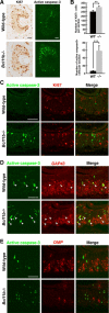
Increased proliferating cells and apoptotic cells in the VNE of Bcl11b−/− mice. A, Proliferative and apoptotic cells were immunostained with anti-Ki67 and anti-active caspase-3 antibodies, respectively. B, The numbers of proliferative and apoptotic cells in the Bcl11b−/− and wild-type VNE were counted. The number of Ki67-positive proliferative cells was slightly increased in Bcl11b−/− mice, and the number of active caspase-3-positive cells was significantly increased in Bcl11b−/− mice. The error bars in B represent the SD of the mean (n = 3, Welch's or Student's t test, **p < 0.01, ***p < 0.005). C, Coronal sections of the VNE at P0 were labeled with an anti-active caspase-3 antibody (green) and an anti-Ki67 antibody (red) in Bcl11b−/− and wild-type mice. No coimmunostaining of Ki67 and active caspase-3 was observed. D, E, Combinations of IHC and ISH were performed to define the step of differentiation when apoptosis occurred in Bcl11b−/− mice. Apoptotic cells were stained using IHC with an anti-active caspase-3 antibody (D, E, green), and immature neurons and mature neurons were detected using ISH with probes for GAP43 (D, red) and OMP (E, red). Double-labeled cells were observed only in immature neurons (D, arrowheads), which indicated that apoptosis predominantly occurred in the immature stages. Scale bars, 50 μm.
Figure 7.
Figure 7.
Changes in gene expression in the VNE of Bcl11b−/− mice. A, B, In situ hybridizations in coronal sections of wild-type (top panels) and Bcl11b−/− (middle panels) VNE at P0, and wild-type VNE at P14 (bottom panels) showed genes with decreased expression (A) or increased expression (B) within the VNE of Bcl11b−/− mice. Each set of panels consists of low-magnification (top) and high-magnification images (bottom). Dashed lines indicate the basal edge of the VNE. Tcfap2e and Meis2 are novel identifiers for the basal and apical VSNs, respectively. Scale bars, 100 μm.
Figure 8.
Figure 8.
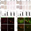
Severely reduced expression of the V1r and V2r genes in the VNE of Bcl11b−/− mice. A, B, The expression of the V1rs and V2rs genes was analyzed using ISH with the following probes: for V1rs: V1rd16 (at E14.5, E16.5, and P0), and V1rb1 and V1re4 (at P0); and for V2rs: V2ra (at E14.5, E16.5 and P0), and V2rb and V2rc (at P0). The lower graphs are quantifications of the number of VR-expressing cells in Bcl11b−/− (gray bar) and wild-type (black bar) mice. The expression of both V1rs and V2rs was significantly reduced in Bcl11b−/− at all of the stages counted. The error bars in A and B represent the SD of the mean (n = 3; Welch's or Student's t test, *p < 0.05, **p < 0.01, ***p < 0.005, ****p < 0.001). C, Apoptosis in VR-expressing VSNs was examined using a combination of ISH and IHC. None of the V1rd16- and V2ra-positive cells (red) overlapped with anti-active caspase-3 immunostaining (green) in Bcl11b−/− and wild-type mice at P0. D, Two-color ISH with the OMP probe (green), and the V1rd16 or the V2ra probe (red) in coronal sections of the VNE showed that VR-expressing VSNs differentiated to maturity in Bcl11b−/− mice (arrowheads indicate coexpression of OMP and VRs). Scale bars, 100 μm.
Figure 9.
Figure 9.
Abnormal development and localization of the two types of VSNs in the VNE of Bcl11b−/− mice. A, ISH with a Gαi2 probe in coronal sections of the Bcl11b−/− and wild-type VNE at E14.5, E16.5, and P0. At E14.5, the expression of Gαi2 was rarely observed in either Bcl11b−/− or wild-type embryos. At E16.5 and P0, Gαi2-expressing cells were increased and were distributed more broadly in the apical–basal axis in the VNE of Bcl11b−/− mice compared with wild-type mice. B, ISH using the Gαo probe in coronal sections of the Bcl11b−/− and wild-type VNE at E14.5, E16.5, and P0. At E14.5, expression of Gαo was detected in the VNE of Bcl11b−/− mice and in wild-type embryos. At E16.5 and P0, the expression of Gαo was decreased in both number and level in the Bcl11b−/− VNE. C, Two-color ISH using the Gαi2 probe (green) and the Gαo (red) probe in coronal sections of the Bcl11b−/− and wild-type VNE at P0. Most Gαi2-positive cells were Gαo positive in both Bcl11b−/− and wild-type mice. Gαi2/Gαo double-positive cells were increased in the VNE of Bcl11b−/− mice, but Gαo single-positive cells were decreased. D, Coexpression of Gαi2 and Gαo were analyzed by two-color ISH in the adult VNE (left: low magnification; right: high magnification of the dotted box). Coexpression of Gαi2 (green) and Gαo (red) were observed in VSNs of the marginal region (typical coexpressing cells are shown in the inset). E, Coexpression of V1rd16 (red) with either Gαi2 or Gαo (green) and coexpression of V2ra (red) with either Gαi2 or Gαo (green) in the wild-type VNE were analyzed using two-color ISH at P0. V1rd16-positive VSNs coexpressed both Gαi2 and Gαo (arrowheads). However, V2ra-positive VSNs coexpressed Gαo (arrowheads), but not Gαi2. Scale bars: 50 μm.
Figure 10.
Figure 10.
A model of the fate determination of vomeronasal sensory neurons. Mash1 is required for the generation of both types of VSNs and acts upstream of Ngn1 and Bcl11b. Two types of neurons are segregated from each other and from the common Gαo-positive postmitotic neurons. The fate of VSN types is regulated by Bcl11b directly or indirectly, and Bcl11b may act as a suppressor for the pathway to the V1r/Gαi2-positive neurons and/or as an activator for the pathway to the V2r/Gαo-positive neurons. In the absence of Bcl11b, normal numbers of neurons are produced because Bcl11b is not essential for neurogenesis in the VSN lineage. However, the loss of Bcl11b function disturbs the fate determination balance between the two types of VSNs, which results in increased V1r/Gαi2-positive neurons and decreased V2r/Gαo-positive neurons.

Comment in

Similar articles

Cited by

References

    1. Albu DI, Feng D, Bhattacharya D, Jenkins NA, Copeland NG, Liu P, Avram D. BCL11B is required for positive selection and survival of double-positive thymocytes. J Exp Med. 2007;204:3003–3015. - PMC - PubMed
    1. Arlotta P, Molyneaux BJ, Chen J, Inoue J, Kominami R, Macklis JD. Neuronal subtype-specific genes that control corticospinal motor neuron development in vivo. Neuron. 2005;45:207–221. - PubMed
    1. Arlotta P, Molyneaux BJ, Jabaudon D, Yoshida Y, Macklis JD. Ctip2 controls the differentiation of medium spiny neurons and the establishment of the cellular architecture of the striatum. J Neurosci. 2008;28:622–632. - PMC - PubMed
    1. Avram D, Fields A, Pretty On Top K, Nevrivy DJ, Ishmael JE, Leid M. Isolation of a novel family of C(2)H(2) zinc finger proteins implicated in transcriptional repression mediated by chicken ovalbumin upstream promoter transcription factor (COUP-TF) orphan nuclear receptors. J Biol Chem. 2000;275:10315–10322. - PMC - PubMed
    1. Berghard A, Buck LB. Sensory transduction in vomeronasal neurons: evidence for G α o, G α i2, and adenylyl cyclase II as major components of a pheromone signaling cascade. J Neurosci. 1996;16:909–918. - PMC - PubMed

Publication types

MeSH terms