. 2011 Jul 13;31(28):10201-5.
doi: 10.1523/JNEUROSCI.0451-11.2011.

Age-related decline in circadian output

Affiliations

Age-related decline in circadian output

Takahiro J Nakamura et al. J Neurosci. .

Abstract

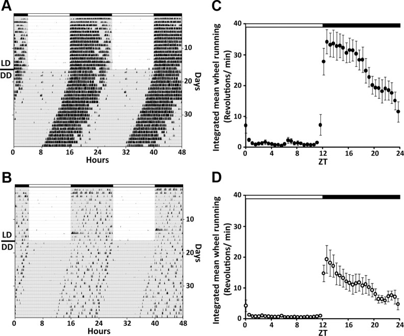
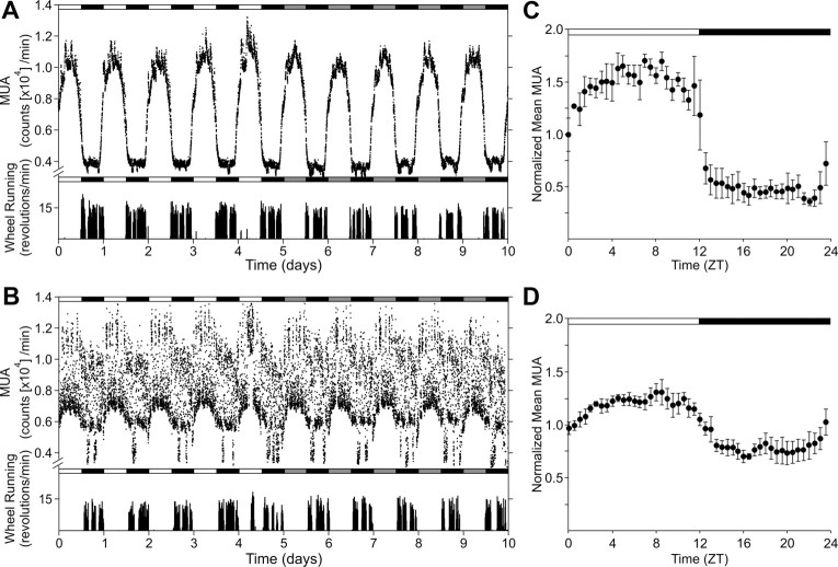
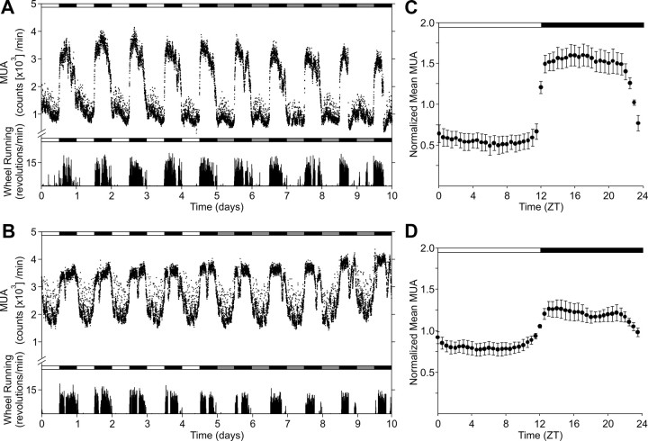
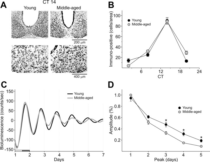
Disruptions in sleep/wake cycles, including decreased amplitude of rhythmic behaviors and fragmentation of the sleep episodes, are commonly associated with aging in humans and other mammals. While there are undoubtedly many factors contributing to these changes, a body of literature is emerging, suggesting that an age-related decline in the central circadian clock in the suprachiasmatic nucleus (SCN) may be a key element responsible. To explore age-related changes in the SCN, we have performed in vivo multiunit neural activity (MUA) recordings from the SCN of freely moving young (3-5 months) and middle-aged (13-18 months) mice. Importantly, the amplitude of day-night difference in MUA was significantly reduced in the older mice. We also found that the neural activity rhythms are clearly degraded in the subparaventricular zone, one of the main neural outputs of the SCN. Surprisingly, parallel studies indicate that the molecular clockwork in the SCN as measured by PER2 exhibited only minor deficits at this same age. Thus, the circadian output measured at the level of neural activity rhythms in the SCN is degraded by aging, and this decline occurs before the disruption of key components of the molecular clockwork.


Figures

Figure 1.
Figure 1.
Reduced circadian behavioral rhythm in middle-aged mice. Representative double-plotted actograms of wheel-running activity in C57BL/6J mice showing the effect of aging on circadian rhythms of locomotor activity of young (A) and middle-aged (B) mice. The mice were maintained in LD cycle for 2 weeks and then transferred into DD. Lighting conditions are indicated at the top of the figure; open bars are light phases and closed are dark. C, D, Integrated mean wheel-running activities were plotted along 24 h for the LD condition (C: young, n = 8; D: middle-aged, n = 10).
Figure 2.
Figure 2.
Reduced MUA rhythm in the SCN of middle-aged mice in vivo. Representative serial-plotted actograms of neural and locomotor activity showing diurnal and circadian rhythms of MUA in the SCN of young (A) and middle-aged (B) mice. Lighting conditions are indicated at the top of the figure; open bars are light phases and closed are dark. Bottom trace represents simultaneous recorded locomotor activity. The number of spikes for MUA or activity counts for locomotor activity was counted every minute. C, D, Integrated mean activities were plotted for the LD condition. Each of four individual recordings was normalized with 24 h moving average and integrated for each group. Data are shown mean ± SEM, n = 4 per group.
Figure 3.
Figure 3.
Reduced MUA rhythm in the SPZ of middle-aged mice in vivo. Representative serial-plotted actograms of neural and locomotor activity showing diurnal rhythms of MUA in the SPZ of young (A) and middle-aged (B) mice. Lighting conditions are indicated at the top of the figure; open bars are light phases and closed are dark. Bottom trace represents simultaneous recorded locomotor activity. The number of spikes for MUA or activity counts for locomotor activity was counted every minute. C, D, Integrated mean activities were plotted for the LD condition. Each of four individual recordings was normalized with 24 h moving average and integrated for each group. Data are shown mean ± SEM, n = 5 per group.
Figure 4.
Figure 4.
Unaltered PER2 expression rhythms in SCN of middle-aged mice. A, Photomicrographs of SCN tissue of each group in middle (200×) and high (400×) magnification. IHC was used to measure PER2 immunoreactivity in the SCN (n = 3–9 per time point) of young and middle-aged mice. Tissue was collected in four phases of the daily cycle (CT 2, 8, 14, 20). B, Numbers of PER2 immunopositive cells in the SCN varied as a function of time of day with highest count in early night (CT 14). C, PER2::LUC rhythms as measured by photomultiplier tube in the SCN. Typical examples of bioluminescence of young and middle-aged SCN explants. D, Amplitude of PER2::LUC expression in the SCN over days in culture. Data are shown mean ± SEM, n = 16 for young group and n = 8 for middle-aged group, *p < 0.05.

References

    1. Abe M, Herzog ED, Yamazaki S, Straume M, Tei H, Sakaki Y, Menaker M, Block GD. Circadian rhythms in isolated brain regions. J Neurosci. 2002;22:350–356. - PMC - PubMed
    1. Asai M, Yoshinobu Y, Kaneko S, Mori A, Nikaido T, Moriya T, Akiyama M, Shibata S. Circadian profile of Per gene mRNA expression in the suprachiasmatic nucleus, paraventricular nucleus, and pineal body of aged rats. J Neurosci Res. 2001;66:1133–1139. - PubMed
    1. Aujard F, Herzog ED, Block GD. Circadian rhythms in firing rate of individual suprachiasmatic nucleus neurons from adult and middle-aged mice. Neurosci. 2001;106:255–261. - PubMed
    1. Biello SM. Circadian clock resetting in the mouse changes with age. Age. 2009;31:293–303. - PMC - PubMed
    1. Bliwise DL. Sleep in normal aging and dementia. Sleep. 1993;16:40–81. - PubMed

Publication types

Substances