

Sleep spindles and hippocampal functional connectivity in human NREM sleep
- PMID: 21753010
- PMCID: PMC6623055
- DOI: 10.1523/JNEUROSCI.5660-10.2011
Sleep spindles and hippocampal functional connectivity in human NREM sleep
Abstract
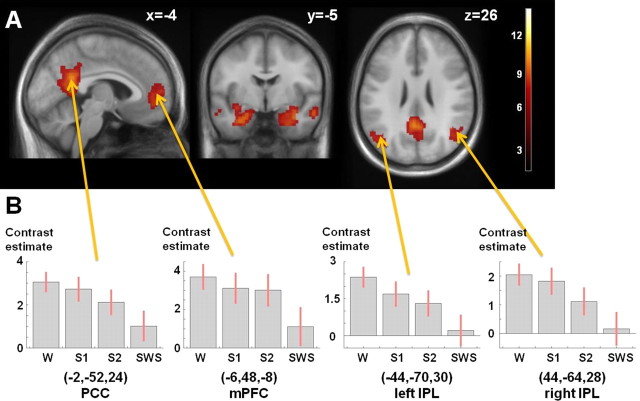
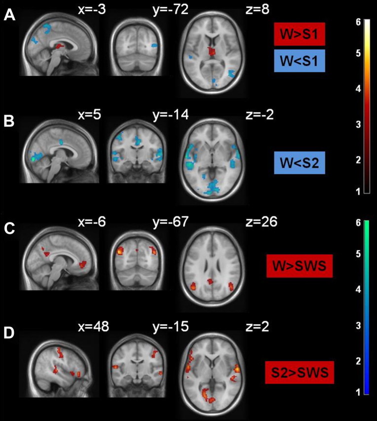
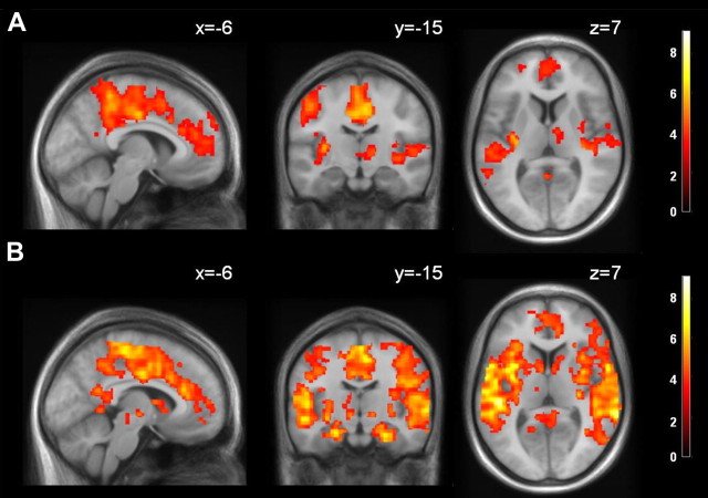
We investigated human hippocampal functional connectivity in wakefulness and throughout non-rapid eye movement sleep. Young healthy subjects underwent simultaneous EEG and functional magnetic resonance imaging (fMRI) measurements at 1.5 T under resting conditions in the descent to deep sleep. Continuous 5 min epochs representing a unique sleep stage (i.e., wakefulness, sleep stages 1 and 2, or slow-wave sleep) were extracted. fMRI time series of subregions of the hippocampal formation (HF) (cornu ammonis, dentate gyrus, and subiculum) were extracted based on cytoarchitectonical probability maps. We observed sleep stage-dependent changes in HF functional coupling. The HF was integrated to variable strength in the default mode network (DMN) in wakefulness and light sleep stages but not in slow-wave sleep. The strongest functional connectivity between the HF and neocortex was observed in sleep stage 2 (compared with both slow-wave sleep and wakefulness). We observed a strong interaction of sleep spindle occurrence and HF functional connectivity in sleep stage 2, with increased HF/neocortical connectivity during spindles. Moreover, the cornu ammonis exhibited strongest functional connectivity with the DMN during wakefulness, while the subiculum dominated hippocampal functional connectivity to frontal brain regions during sleep stage 2. Increased connectivity between HF and neocortical regions in sleep stage 2 suggests an increased capacity for possible global information transfer, while connectivity in slow-wave sleep is reflecting a functional system optimal for segregated information reprocessing. Our data may be relevant to differentiating sleep stage-specific contributions to neural plasticity as proposed in sleep-dependent memory consolidation.
Figures
References
-
- Amunts K, Kedo O, Kindler M, Pieperhoff P, Mohlberg H, Shah NJ, Habel U, Schneider F, Zilles K. Cytoarchitectonic mapping of the human amygdala, hippocampal region and entorhinal cortex: intersubject variability and probability maps. Anat Embryol (Berl) 2005;210:343–352. - PubMed
-
- Axmacher N, Mormann F, Fernández G, Elger CE, Fell J. Memory formation by neuronal synchronization. Brain Res Rev. 2006;52:170–182. - PubMed
Publication types
MeSH terms
LinkOut - more resources
Full Text Sources
Research Materials
Miscellaneous