. 2011 Jul 13;31(28):10392-402.
doi: 10.1523/JNEUROSCI.0595-11.2011.

Rules ventral prefrontal cortical axons use to reach their targets: implications for diffusion tensor imaging tractography and deep brain stimulation for psychiatric illness

Affiliations

Rules ventral prefrontal cortical axons use to reach their targets: implications for diffusion tensor imaging tractography and deep brain stimulation for psychiatric illness

Julia F Lehman et al. J Neurosci. .

Abstract

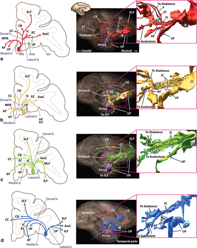
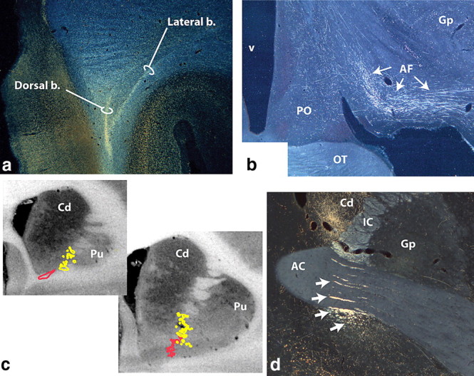
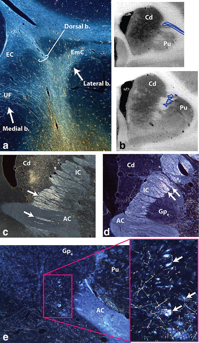
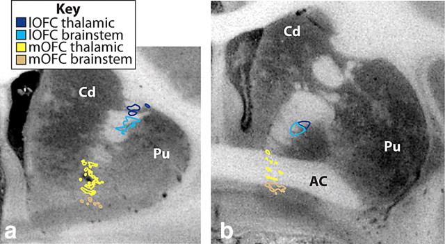
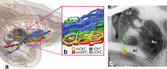
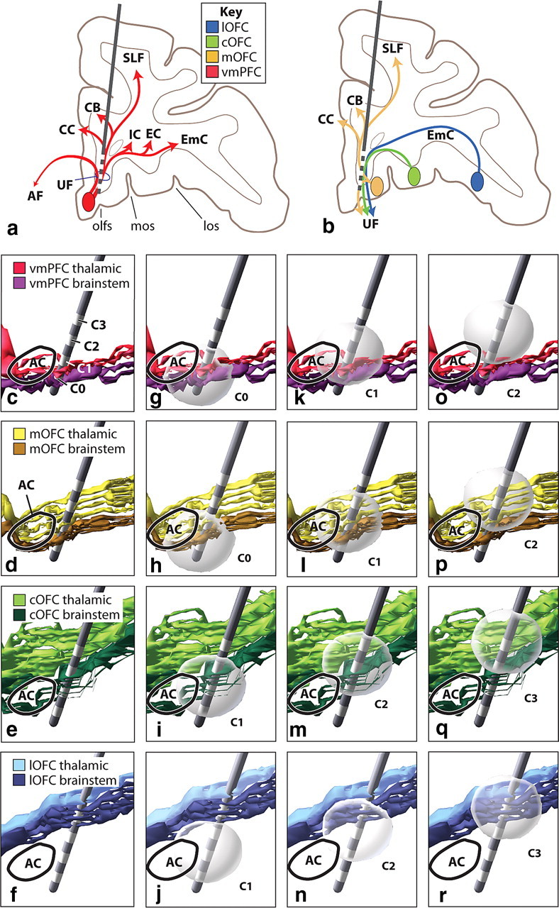
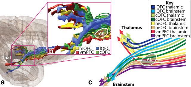
The ventral prefrontal cortex (vPFC) is involved in reinforcement-based learning and is associated with depression, obsessive-compulsive disorder, and addiction. Neuroimaging is increasingly used to develop models of vPFC connections, to examine white matter (WM) integrity, and to target surgical interventions, including deep brain stimulation. We used primate (Macaca nemestrina/Macaca fascicularis) tracing studies and 3D reconstructions of WM tracts to delineate the rules vPFC projections follow to reach their targets. vPFC efferent axons travel through the uncinate fasciculus, connecting different vPFC regions and linking different functional regions. The uncinate fasciculus also is a conduit for vPFC fibers to reach other cortical bundles. Fibers in the internal capsule are organized according to destination. Thalamic fibers from each vPFC region travel dorsal to their brainstem fibers. The results show regional differences in the trajectories of fibers from different vPFC areas. Overall, the medial/lateral vPFC position dictates the route that fibers take to enter major WM tracts, as well as the position within specific tracts: axons from medial vPFC regions travel ventral to those from more lateral areas. This arrangement, coupled with dorsal/ventral organization of thalamic/brainstem fibers through the internal capsule, results in a complex mingling of thalamic and brainstem axons from different vPFC areas. Together, these data provide the foundation for dividing vPFC WM bundles into functional components and for predicting what is likely to be carried at different points through each bundle. These results also help determine the specific connections that are likely to be captured at different neurosurgical targets.


Figures

Figure 1.
Figure 1.
vPFC fiber pathways. a, vmPFC. b, mOFC. c, cOFC. d, lOFC. Left panels illustrate how different bundles separate from the injection site as they enter the white matter. Note, in each case, fibers divide into medial, dorsal, and lateral pathways (blue). However, the specific bundles that are carried within each depend on the position of the origin of the fibers. Right panels illustrate 3D renderings of a lateral view of a sagittal plane. Each is accompanied with an inset to better visualize the separation of fiber bundles. External and extreme capsule pathways have been removed for clarity. Asterisks indicate the rostrocaudal position of the injection site. AC indicates location of the anterior commissure. Axons from the vPFC, mOFC, and cOFC travel ventral or through the AC. Note that axons traveling through the internal capsule divide into dorsal thalamic fibers and ventral brainstem axons. Pathways traveling to the temporal lobe (uncinate fasciculus and ventral amygdalofugal bundle) primarily arise from separate bundles. AF, Ventral amygdalofugal pathway; Amyg, amygdala; b., bundle; CB, cingulum bundle; CC, corpus callosum; EC, external capsule; EmC, extreme capsule; F, fornix; IC, internal capsule; ILF, inferior longitudinal fasciculus; los, lateral orbital sulcus; MFB, medial forebrain bundle; MLF, middle longitudinal fasciculus; mos, medial orbital sulcus; olfs, olfactory sulcus; SLF, superior longitudinal fasciculus; ST, stria terminalis; UF, uncinate fasciculus; *injection site location.
Figure 2.
Figure 2.
Photomicrographs and schematics of vmPFC and mOFC pathways. a, vmPFC axons leave the injection site traveling dorsally and divide into the dorsal and lateral bundles. Some dorsal fibers enter the emerging corpus callosum. The medial bundle remains ventral (not illustrated). b, The ventral amygdalofugal pathway carries fibers from the vmPFC to the amygdala. c, Fibers from the vmPFC (red) and mOFC (yellow) enter the IC ventrally and form fascicules within the ventral striatum. d, Photomicrograph illustrating mOFC IC axons embedded within the AC or ventral to it (arrows). Those within the IC terminate in the thalamus; the ventral groups continue to the brainstem. AC, Anterior commissure; AF, ventral amygdalofugal pathway; b., bundle; Cd, caudate nucleus; Gp, globus pallidus; IC, internal capsule; OT, optic tract; PO, preoptic area; Pu, putamen; v, ventricle.
Figure 3.
Figure 3.
Photomicrographs of cOFC and lOFC pathways. a, lOFC fibers dividing into medial (UF), dorsal, and lateral bundles. Fibers in the dorsal bundle pass directly through the frontal white matter to reach the corpus callosum and superior longitudinal fasciculus or enter the external or extreme capsules. b, Fibers from the lOFC enter the IC dorsolaterally. c, Fibers from the cOFC pass through the anterior commissure to the brainstem; those dorsal to the commissure terminate in the thalamus. d, All descending IC lOFC axons are located dorsal to the anterior commissure. Note the relative lateral position compared with the cOFC fibers. e, Axons from the lOFC form small fascicles (arrows) that leave the external capsule travel medial and enter the amygdalofugal pathway or terminate in the hypothalamus. AC, Anterior commissure; b., bundle; Cd, caudate nucleus; EC, external capsule; EmC, extreme capsule; Gpe, globus pallidus, external segment; IC, internal capsule; Pu, putamen; UF, uncinate fasciculus.
Figure 4.
Figure 4.
Schematic and photomicrographs illustrating the complexity of the uncinate fasciculus. a, Schematic illustrating vPFC axons crossing through the UF to reach other fiber tracts. Note that the medial and central parts of the UF have more crossing fibers compared with the lateral part of the tract. bc, Axons from a cOFC injection site (*) enter the UF (b), travel a few millimeters and split into different bundles (c). Note the crossing of many fibers as the axons enter the UF in b (arrows). b., Bundle; CB, cingulum bundle; CC, corpus callosum; EC, external capsule; EmC, extreme capsule; IC, internal capsule; los, lateral orbital sulcus; mos, medial orbital sulcus; UF, uncinate fasciculus.
Figure 5.
Figure 5.
Coronal sections illustrating the different positions of thalamic versus brainstem mOFC fibers (yellow–tan) and lOFC (dark blue–light blue) entering and traveling through the IC. Brainstem fibers (tan and light blue) travel ventral to thalamic fibers (yellow and dark blue). AC, Anterior commissure; Cd, caudate nucleus; lOFC, lateral orbital frontal cortex; mOFC, medial orbital frontal cortex; Pu, putamen.
Figure 6.
Figure 6.
vPFC fibers through internal capsule. a, Overview of the internal capsule in the parasagittal plane. b, Enlargement of the anterior internal capsule showing the dorsal/ventral topography rostral to the AC. Note the medial/lateral topography, with medial vPFC fibers traveling ventral to lateral vPFC axons. c, Coronal section illustrating the organization of the vPFC fibers in the IC at the level of the anterior commissure. AC, Anterior commissure; Cd, caudate nucleus; cOFC, central orbital frontal cortex; lOFC, lateral orbital frontal cortex; mOFC, medial orbital frontal cortex; Pu, putamen; Thal, thalamus; vmPFC, ventral medial prefrontal cortex.
Figure 7.
Figure 7.
Organization of vPFC pathways in the corpus callosum. a, Sagittal view of 3D model of vPFC pathways in corpus callosum. Fibers originating from more lateral vPFC regions (blue) cross dorsally to fibers from more medial vPFC areas (red). b, Micrograph of cOFC fibers crossing mid-level genu of corpus callosum. c, Micrograph of vmPFC crossing most ventral portion of corpus callosum. GCC, Genu of corpus callosum; LV, lateral ventricle; blue, lOFC; green, cOFC; red, vmPFC.
Figure 8.
Figure 8.
Modeled DBS electrodes at the SCGwm and VC/VS targets. a, The SCGwm target involves all fibers from cortical areas adjacent to the electrode, including descending projections. b, SCGwm target also involves other vPFC fibers that pass through the site, including axons from lateral vPFC regions traveling medially and those from medial OFC areas traveling dorsal. cf, Sagittal view of specific ventral PFC bundles traveling in the IC with an electrode representation embedded at the VC/VS site. Each contact captures a different set of thalamic and/or brainstem fibers. gr, Spheres indicating volume of tissue activated by electrical stimulation of a pulse width of 60 μs and 3V at each contact site. The electrode field model is not more oval shaped because we used the isotropic simplification that allowed us to move the electrode to any position. AF, Ventral amygdalofugal bundle; AC, anterior commissure; C0–C3, contacts 0–3; CB, cingulum bundle; CC, corpus callosum; cOFC, central orbital prefrontal cortex; EC, external capsule; EmC, extreme capsule; IC, internal capsule; lOFC, lateral orbital prefrontal cortex; los, lateral orbital sulcus; mOFC, medial orbital prefrontal cortex; mos, medial orbital sulcus; olfs, olfactory sulcus; SLF, superior longitudinal fasciculus; UF, uncinate fasciculus; vmPFC, ventral medial prefrontal cortex.
Figure 9.
Figure 9.
White matter fascicules through the ventral forebrain. a, Nissl-stained sagittal section through the monkey brain. Arrows indicate white matter tracts from the vPFC. bc, Acetylcholinesterase-stained coronal sections through the human striatum at different rostral/caudal levels. Arrows indicate WM fascicules passing through the ventral striatum. AC, Anterior commissure; Cd, caudate; IC, internal capsule; Pu, putamen. Asterisks indicate blood vessels.
Figure 10.
Figure 10.
A parasagittal section illustrating the relationship between brainstem and thalamic internal capsule fibers from the different vPFC regions. a, Overview of vPFC fibers in the IC. b, Enlargement of the anterior internal capsule showing the split of thalamic and brainstem fibers caudal to the anterior commissure. c, Schematic of the separation of thalamic and brainstem fibers. Thalamic fibers from vmPFC (red) occupy a similar position as brainstem fibers from mOFC (orange); mOFC thalamic fibers (yellow) occupy a similar position as brainstem fibers from cOFC (dark green). AC, Anterior commissure; cOFC, central orbital frontal cortex; mOFC, medial orbital frontal cortex; lOFC, lateral orbital frontal cortex; Thal, thalamus; vmPFC, ventral medial prefrontal cortex.

References

    1. Amaral DG, Price JL, Pitkanen A, Carmichael ST. Anatomical organization of the primate amygdaloid complex. In: Aggleton JP, editor. The amygdala: neurobiological aspects of emotion, memory, and mental dysfunction. New York: Wiley; 1992. pp. 1–66.
    1. Beckmann M, Johansen-Berg H, Rushworth MF. Connectivity-based parcellation of human cingulate cortex and its relation to functional specialization. J Neurosci. 2009;29:1175–1190. - PMC - PubMed
    1. Beevor CE, Horsley V. An experimental investigation into the arrangement of the excitable fibres of the internal capsule of the bonnet monkey (Macacus sinicus) Philos Trans R Soc Lond B Biol Sci. 1890;181:49–88.
    1. Behrens TE, Berg HJ, Jbabdi S, Rushworth MF, Woolrich MW. Probabilistic diffusion tractography with multiple fibre orientations: What can we gain? Neuroimage. 2007;34:144–155. - PMC - PubMed
    1. Browning PG, Gaffan D. Impairment in object-in-place scene learning after uncinate fascicle section in macaque monkeys. Behav Neurosci. 2008;122:477–482. - PubMed

Publication types

LinkOut - more resources