Review
. 2011 Oct;301(4):F684-96.
doi: 10.1152/ajprenal.00259.2011. Epub 2011 Jul 13.

ENaC structure and function in the wake of a resolved structure of a family member

Affiliations
Review

ENaC structure and function in the wake of a resolved structure of a family member

Ossama B Kashlan et al. Am J Physiol Renal Physiol. 2011 Oct.

Abstract

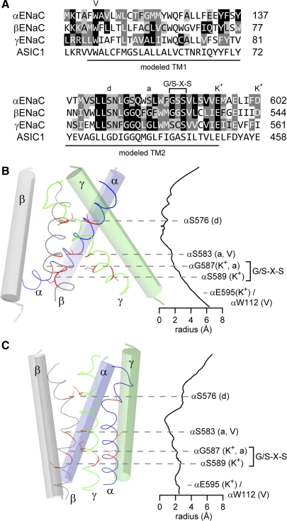
Our understanding of epithelial Na(+) channel (ENaC) structure and function has been profoundly impacted by the resolved structure of the homologous acid-sensing ion channel 1 (ASIC1). The structure of the extracellular and pore regions provide insight into channel assembly, processing, and the ability of these channels to sense the external environment. The absence of intracellular structures precludes insight into important interactions with intracellular factors that regulate trafficking and function. The primary sequences of ASIC1 and ENaC subunits are well conserved within the regions that are within or in close proximity to the plasma membrane, but poorly conserved in peripheral domains that may functionally differentiate family members. This review examines functional data, including ion selectivity, gating, and amiloride block, in light of the resolved ASIC1 structure.


Figures

Fig. 1.
Fig. 1.
Acid-sensing ion channel 1 (ASIC1) structure. A: ribbon diagram of ASIC1 trimer (pdb code: 3HGC)*. One subunit is shown as a solid ribbon, with domains in the extracellular region indicated. The other 2 subunits are shown as transparent ribbons. The approximate positions of the outer (Out) and inner (In) borders of the cell membrane are indicated. B: topology diagram of ASIC1. α-Helices and β-strands are represented by red cylinders and blue arrows, respectively. Labeling of secondary structure elements are as in Jasti et al. (63). This figure was adapted from a figure originally published in J Biol Chem 285: 35216–35223, 2010.
Fig. 2.
Fig. 2.
Epithelial Na+ channel (ENaC) pore comparative models. A: pore sequence alignment between chicken ASIC1 and mouse ENaC subunits. Identity (black background, white type), strong similarity (dark gray background, white type), and weak similarity (light gray background) are indicated. Comparative models of sequences indicated in A were generated using MODELLER (49), a clockwise α-γ-β-subunit arrangement, and either the 3HGC (B) or 2QTS (C) ASIC1 pdb coordinates. Seven different models are possible given this set of constraints: 1 for the symmetric 3HGC structure and 6 for the asymmetric 2QTS structure that presented 2 slightly different ASIC1 trimers (1.6 Å rmsd for pore Cα). For the sake of brevity, we made one model based on the 2QTS structure using the A, B, and C chains, and aligned the ENaC β-subunit to chain A based on previous work suggesting that the TM2 of the β-subunit is shifted toward the cytosol relative to the α- and γ-ENaC subunits (88). B and C: pore radius along the pore axis was calculated using HOLE (126). The Gly/Ser-X-Ser selectivity tract and mutations at select sites that affect ENaC open probability (Po), degenerin site (d), amiloride block (a), voltage sensitivity (V), and Na+/K+ selectivity (K+) are indicated. B and C: select sites are highlighted with dashed lines indicating the approximate level of homologous α, β, and γ residues in the pore.
Fig. 3.
Fig. 3.
ENaC α-subunit model highlighting proposed inhibitory peptide and Cl binding sites. Shown is α trimer, with 1 subunit represented as a ribbon, and the other 2 subunits represented as a surface with relatively basic (blue) and acidic (red) areas indicated. Positions of bound Cl (green spheres) were derived from the ASIC1 structure (pdb code: 3HGC). Ribbon is colored by domain: red, TM helices; yellow, palm, β-ball, and knuckle; green, finger; orange, thumb. Five disulfide bonds in the thumb are highlighted (orange spheres). The 8 inhibitory tract residues essential for peptide inhibition are highlighted (purple spheres), while the remaining residues of the furin-excised inhibitory tract are removed.

References

    1. Abi-Antoun T, Shi S, Tolino LA, Kleyman TR, Carattino MD. Second transmembrane domain modulates epithelial sodium channel gating in response to shear stress. Am J Physiol Renal Physiol 300: F1089–F1095, 2011 - PMC - PubMed
    1. Adachi M, Kitamura K, Miyoshi T, Narikiyo T, Iwashita K, Shiraishi N, Nonoguchi H, Tomita K. Activation of epithelial sodium channels by prostasin in Xenopus oocytes. J Am Soc Nephrol 12: 1114–1121, 2001 - PubMed
    1. Adams CM, Anderson MG, Motto DG, Price MP, Johnson WA, Welsh MJ. Ripped pocket and pickpocket, novel Drosophila DEG/ENaC subunits expressed in early development and in mechanosensory neurons. J Cell Biol 140: 143–152, 1998 - PMC - PubMed
    1. Adams CM, Snyder PM, Welsh MJ. Paradoxical stimulation of a DEG/ENaC channel by amiloride. J Biol Chem 274: 15500–15504, 1999 - PubMed
    1. Adebamiro A, Cheng Y, Rao US, Danahay H, Bridges RJ. A segment of γ ENaC mediates elastase activation of Na+ transport. J Gen Physiol 130: 611–629, 2007 - PMC - PubMed

Publication types

MeSH terms

LinkOut - more resources