. 2011 Aug 15;187(4):1845-55.
doi: 10.4049/jimmunol.1100377. Epub 2011 Jul 13.

IFN-β mediates suppression of IL-12p40 in human dendritic cells following infection with virulent Francisella tularensis

Affiliations

IFN-β mediates suppression of IL-12p40 in human dendritic cells following infection with virulent Francisella tularensis

Timothy J Bauler et al. J Immunol. .

Abstract

Active suppression of inflammation is a strategy used by many viral and bacterial pathogens, including virulent strains of the bacterium Francisella tularensis, to enable colonization and infection in susceptible hosts. In this study, we demonstrated that virulent F. tularensis strain SchuS4 selectively inhibits production of IL-12p40 in primary human cells via induction of IFN-β. In contrast to the attenuated live vaccine strain, infection of human dendritic cells with virulent SchuS4 failed to induce production of many cytokines associated with inflammation (e.g., TNF-α and IL-12p40). Furthermore, SchuS4 actively suppressed secretion of these cytokines. Assessment of changes in the expression of host genes associated with suppression of inflammatory responses revealed that SchuS4, but not live vaccine strain, induced IFN-β following infection of human dendritic cells. Phagocytosis of SchuS4 and endosomal acidification were required for induction of IFN-β. Further, using a defined mutant of SchuS4, we demonstrated that the presence of bacteria in the cytosol was required, but not sufficient, for induction of IFN-β. Surprisingly, unlike previous reports, induction of IFN-β by F. tularensis was not required for activation of the inflammasome, was not associated with exacerbation of inflammatory responses, and did not control SchuS4 replication when added exogenously. Rather, IFN-β selectively suppressed the ability of SchuS4-infected dendritic cells to produce IL-12p40. Together, these data demonstrated a novel mechanism by which virulent bacteria, in contrast to attenuated strains, modulate human cells to cause disease.


Figures

Figure 1
Figure 1
Differential induction of pro-inflammatory cytokines by attenuated and virulent strains of F. tularensis. Primary human monocyte-derived DC (hDC) were infected with a MOI=50 with the indicated strains of F. tularensis. (A) Intracellular bacteria were enumerated at the indicated times post infection. *=p<0.01 compared to SchuS4 infected hDC. (B) Supernatants were harvested from uninfected or F. tularensis infected cultures at the indicated time points after infection and analyzed for TNF-α, IL-6, and IL-12p40 by ELISA. hDC stimulated 24 hours prior to harvest with ultrapure E. coli LPS (10 ng/ml) served as positive controls. *=p<0.01 compared to uninfected and SchuS4 infected hDC. ** = p<0.001 compared to all samples. (C) SchuS4 or mock infected hDC cultures were stimulated 24 hours after infection with ultrapure E. coli LPS. Concentrations of TNF-α, IL-6, and IL-12p40 in culture supernatants were determined a further 24 hours after LPS treatment. *=p<0.01 compared to uninfected, LPS treated samples. Error bars represent SEM. Each data point represents the mean of triplicate samples. Panel A is the mean of 8 different experiments and Panel B and C are representative of 3 experiments of similar design.
Figure 2
Figure 2
Induction of IFN-β by virulent F. tularensis. (A) hDC were infected with SchuS4. RNA was harvested at the indicated time points after infection for analysis of host genes by qRT-PCR. The fold change of TNF-α, IL-1β, IL-10, Tollip, IFN-α, IFN-β and IFN-γ, normalized to uninfected samples, are depicted. (B) RNA from hDC infected with F. tularensis strain SchuS4 or LVS was harvested at the indicated time points after infection for analysis of IFN-β by qRT-PCR. IFN-β transcript levels were normalized to those from uninfected hDC. The intracellular bacterial loads at the indicated time points are denoted. *= p<0.01 compared to LVS infected cells at each time point. (C) hDC were infected with SchuS4 or LVS and 12 hours after infection culture supernatants were for IFN-β by ELISA. Uninfected hDC served as negative controls. * = p<0.05 compared to LVS and uninfected samples. Error bars represent SEM. Each data point represents the mean of triplicate samples. Data are representative 3 experiments of similar design.
Figure 3
Figure 3
Requirements for SchuS4-mediated IFN-β production in hDC. One hour prior to infection, hDC were treated with (A) cytochalasin D to inhibit phagocytosis or (B) bafilomycin A to inhibit endosomal acidification. Eight hours after infection, RNA was extracted for analysis by qRT-PCR. RNA collected from hDC treated with LPS served as a positive control. IFN-β transcript levels were normalized to those from mock-infected cells. *=p<0.01 compared to untreated, SchuS4 infected hDC. **=p<0.01 compared to hDC treated with LPS in the absence of inhibitors. (C) hDC were exposed to SchuS4 that had been killed with 2% PFA (SchuS4+PFA). Eight hours after infection, RNA was extracted for analysis of IFN-β transcripts by qRT-PCR. *=p<0.01 compared to PFA killed SchuS4. Error bars represent SEM. Each data point represents the mean of triplicate samples. Data are representative of 3 experiments of similar design.
Figure 4
Figure 4
Endosomal escape and early cytosolic replication are not sufficient for SchuS4-mediated induction of IFN-β. hDC were infected with the indicated SchuS4 strains. (A) Intracellular bacteria were enumerated at the indicated times points after infection. (B) Cytoplasmic bacteria were identified 3 hours after infection using a phagosomal integrity assay and were enumerated by microscopy. (C) RNA was harvested 8 hours after infection for assessment of IFN-β transcript by qRT-PCR. IFN-β transcript levels were normalized to those from uninfected hDC. *=p<0.05 compared to all other samples. Error bars represent SEM. Each data point represents the mean of triplicate samples. Panels A and C represent the mean of 5 independent experiments, and data in panel B is representative of 3 experiments of similar design.
Figure 5
Figure 5
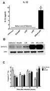
IFN-β is not correlated with activation of the inflammasome following F. tularensis infection of hDC. (A–B) hDC were mock infected or infected with the indicated strains of F. tularensis, or treated with E. coli LPS with or without pretreatment with rhIFN-β. (A) Forty-eight hours after infection supernatants were collected and analyzed for IL-1β by ELISA. * = p<0.0001 compared to all other samples. (B) Eight hours after infection intracellular pro-IL-1β was detected in hDC lysates by Western blotting. Blots were stripped and reprobed with anti-β-actin to demonstrate equal loading. (C) hDC were mock infected or infected with F. tularensis SchuS4 or LVS. At the indicated time points after infection, the number of hDC positive for activated caspase-1 was detected by flow cytometry using Green FLICA Caspase-1 assay kit. *=p<0.05 compared to all other samples. **=p<0.01 compared to all other samples. Error bars represent SEM. Each data point represents the mean of triplicate samples. Data are representative of 3 experiments of similar design.
Figure 6
Figure 6
SchuS4 induced IFN-β selectively inhibits IL-12p40. (A) hDC were treated with PBS (−) or the indicated concentration of rhIFN-β for 16 hours prior to exposure to E. coli LPS. Supernatants were harvested 24 hours later and examined for IL-12p40 and TNF-α by ELISA. * = p<0.01 compared to cells treated with LPS alone. (B) hDC were infected with SchuS4 in the presence of neutralizing polyclonal anti-IFN-β antibody or polyclonal rabbit IgG (isotype) control. Sixteen hours later, infected or mock infected hDC were treated with ultrapure E. coli LPS and assessed for intracellular IL-12p40 and TNF-α by flow cytometry. Data was normalized by defining the percentage of cells in an uninfected culture of hDC that respond to LPS by production of cytokine as 100%. Significance is indicated as the p value on each graph. ns = not significant. (C) hDC were infected with SchuS4 in the presence of neutralizing monoclonal anti-IFN-β antibody or mouse IgG1 (isotype) control. Sixteen hours later, infected or mock infected hDC were treated with ultrapure E. coli LPS. Sixteen hours later culture supernatants were assessed for IL-12p40 and TNF-α by ELISA. Data was normalized by defining the concentration of cytokine secretion in an uninfected culture of hDC that respond to LPS by production of cytokine as 100%. Significance is indicated as the p value on each graph. ns = not significant. (D) hDC were infected with the indicated SchuS4 strains. Uninfected hDC served as negative controls. Supernatants were harvested 48 hours after infection and analyzed for IL-12p40 and TNF-α by ELISA. *=p<0.05 compared to all other samples. (E) hDC were treated with rhIFN-β and infected with LVS. Twenty-four hours after infection supernatant were assessed for IL-12p40 by ELISA. Uninfected hDC served as negative controls. * = p<0.01 compared to untreated, LVS infected hDC. (F) hDC were primed with rhIFN-β followed by infection with the designated strains of F. tularensis. Intracellular bacteria were enumerated at 3 and 24 hours after infection. *=p<0.001 compared to all other samples. Each data point represents the mean of triplicate samples. Error bars represent SEM. Data are representative of 3 experiments of similar design.

Similar articles

Cited by

References

    1. Coburn B, Li Y, Owen D, Vallance BA, Finlay BB. Salmonella enterica serovar Typhimurium pathogenicity island 2 is necessary for complete virulence in a mouse model of infectious enterocolitis. Infect Immun. 2005;73:3219–3227. - PMC - PubMed
    1. Faure K, Sawa T, Ajayi T, Fujimoto J, Moriyama K, Shime N, Wiener-Kronish JP. TLR4 signaling is essential for survival in acute lung injury induced by virulent Pseudomonas aeruginosa secreting type III secretory toxins. Respir Res. 2004;5:1. - PMC - PubMed
    1. Bosio CM, Aman MJ, Grogan C, Hogan R, Ruthel G, Negley D, Mohamadzadeh M, Bavari S, Schmaljohn A. Ebola and Marburg viruses replicate in monocyte-derived dendritic cells without inducing the production of cytokines and full maturation. J Infect Dis. 2003;188:1630–1638. - PubMed
    1. Bosio CM, Bielefeldt-Ohmann H, Belisle JT. Active suppression of the pulmonary immune response by Francisella tularensis Schu4. J Immunol. 2007;178:4538–4547. - PubMed
    1. Salcedo SP, Marchesini MI, Lelouard H, Fugier E, Jolly G, Balor S, Muller A, Lapaque N, Demaria O, Alexopoulou L, Comerci DJ, Ugalde RA, Pierre P, Gorvel JP. Brucella control of dendritic cell maturation is dependent on the TIR-containing protein Btp1. PLoS Pathog. 2008;4:e21. - PMC - PubMed

Publication types

MeSH terms