

Adherens junction proteins in the hamster uterus: their contributions to the success of implantation
- PMID: 21753191
- PMCID: PMC3197917
- DOI: 10.1095/biolreprod.110.090126
Adherens junction proteins in the hamster uterus: their contributions to the success of implantation
Abstract
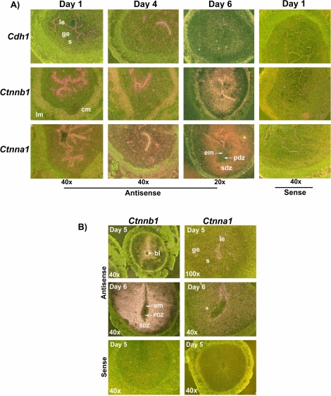
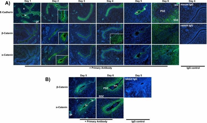
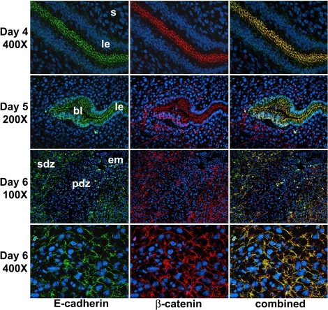
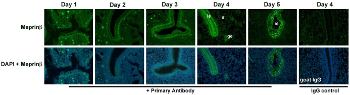
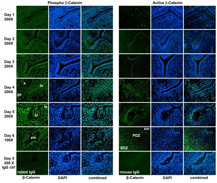
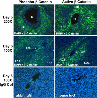
The adherens junction (AJ) is important for maintaining uterine structural integrity, composition of the luminal environment, and initiation of implantation by virtue of its properties of cell-cell recognition, adhesion, and establishment of cell polarity and permeability barriers. In this study, we investigated the uterine changes of AJ components E-cadherin, beta-catenin, and alpha-catenin at their mRNA and protein levels, together with the cellular distribution of meprinbeta, phospho-beta-catenin, and active beta-catenin proteins, in hamsters that show only ovarian progesterone-dependent uterine receptivity and implantation. By in situ hybridization and immunofluorescence, we have demonstrated that uterine epithelial cells expressed three of these AJ proteins and their mRNAs prior to and during the initial phase of implantation. Immunofluorescence study showed no change in epithelial expression patterns of uterine AJ proteins from Days 1 to 5 of pregnancy. With advancement of the implantation process, AJ components were primarily expressed in cells of the secondary decidual zone (SDZ), but not in the primary decidual zone (PDZ). In contrast, we noted strong expression of beta-catenin and alpha-catenin proteins in the PDZ, but not in the SDZ, of mice. Taken together, these results suggest that AJ proteins contribute to uterine barrier functions by cell-cell adhesion to ensure protection of the embryo. In addition, cleavage of E-cadherin by meprinbeta might contribute to weakening uterine epithelial cell-cell contact for blastocyst implantation. We also report that the nuclear localization of active beta-catenin from Day 4 onward in hamsters implies that beta-catenin/Wnt-signal transduction is activated in the uterus during implantation and decidualization.
Figures
Similar articles
-
Leukemia inhibitory factor ligand-receptor signaling is important for uterine receptivity and implantation in golden hamsters (Mesocricetus auratus).Reproduction. 2008 Jan;135(1):41-53. doi: 10.1530/REP-07-0013. Reproduction. 2008. PMID: 18159082
-
Tight and adherens junctions in the ovine uterus: differential regulation by pregnancy and progesterone.Endocrinology. 2007 Aug;148(8):3922-31. doi: 10.1210/en.2007-0321. Epub 2007 May 3. Endocrinology. 2007. PMID: 17478549
-
Zonula occludens-1 and E-cadherin are coordinately expressed in the mouse uterus with the initiation of implantation and decidualization.Dev Biol. 1999 Apr 15;208(2):488-501. doi: 10.1006/dbio.1999.9206. Dev Biol. 1999. PMID: 10191061
-
Dynamics of adherens junctions in epithelial establishment, maintenance, and remodeling.J Cell Biol. 2011 Mar 21;192(6):907-17. doi: 10.1083/jcb.201009141. J Cell Biol. 2011. PMID: 21422226 Free PMC article. Review.
-
Embryo-uterine cross-talk during implantation: the role of Wnt signaling.Mol Hum Reprod. 2009 Apr;15(4):215-21. doi: 10.1093/molehr/gap009. Epub 2009 Feb 17. Mol Hum Reprod. 2009. PMID: 19223336 Review.
Cited by
-
Egr1 protein acts downstream of estrogen-leukemia inhibitory factor (LIF)-STAT3 pathway and plays a role during implantation through targeting Wnt4.J Biol Chem. 2014 Aug 22;289(34):23534-45. doi: 10.1074/jbc.M114.588897. Epub 2014 Jul 10. J Biol Chem. 2014. PMID: 25012664 Free PMC article.
-
miRNA profiling in intrauterine exosomes of pregnant cattle on day 7.Front Vet Sci. 2022 Dec 20;9:1078394. doi: 10.3389/fvets.2022.1078394. eCollection 2022. Front Vet Sci. 2022. PMID: 36605764 Free PMC article.
-
The immunolocalization of cadherins and beta-catenin in the cervix and vagina of cycling cows.Vet Res Commun. 2023 Sep;47(3):1155-1175. doi: 10.1007/s11259-023-10075-4. Epub 2023 Feb 2. Vet Res Commun. 2023. PMID: 36729278
-
Adrenomedullin improves fertility and promotes pinopodes and cell junctions in the peri-implantation endometrium.Biol Reprod. 2017 Sep 1;97(3):466-477. doi: 10.1093/biolre/iox101. Biol Reprod. 2017. PMID: 29025060 Free PMC article.
-
Estrogen Regulates the Expression and Localization of YAP in the Uterus of Mice.Int J Mol Sci. 2022 Aug 29;23(17):9772. doi: 10.3390/ijms23179772. Int J Mol Sci. 2022. PMID: 36077170 Free PMC article.
References
-
- Huet-Hudson YM, Andrews GK, Dey SK. Cell type-specific localization of c-myc protein in the mouse uterus: modulation by steroid hormones and analysis of the periimplantation period. Endocrinology 1989; 125: 1683 1690. - PubMed
-
- Kemler R, Ozawa M, Ringwald M. Calcium-dependent cell adhesion molecules. Curr Opin Cell Biol 1989; 1: 892 897. - PubMed
Publication types
MeSH terms
Substances
Grants and funding
LinkOut - more resources
Full Text Sources
