

Sex differences in cardiomyocyte connexin43 expression
- PMID: 21753256
- PMCID: PMC3136750
- DOI: 10.1097/FJC.0b013e31821b70b4
Sex differences in cardiomyocyte connexin43 expression
Abstract
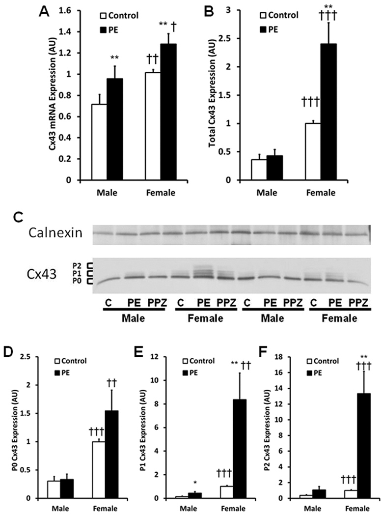
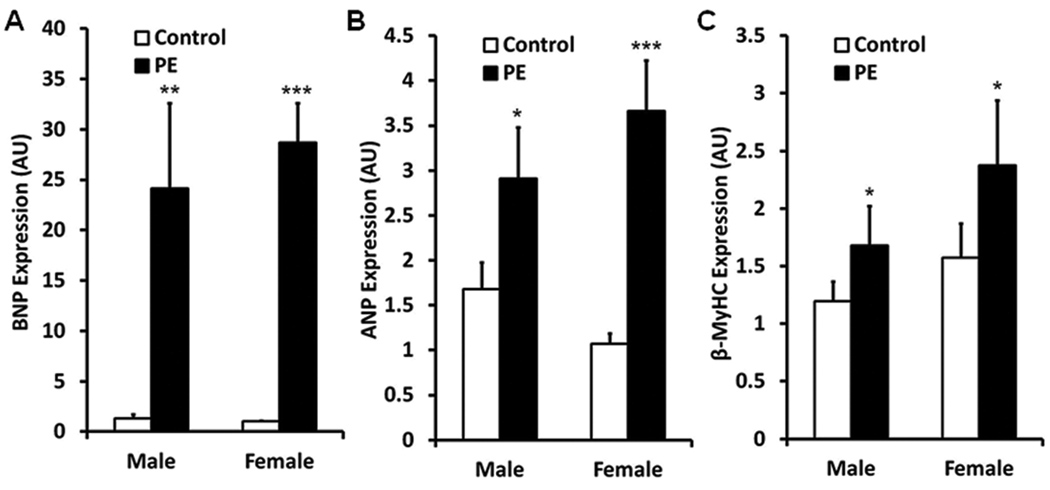
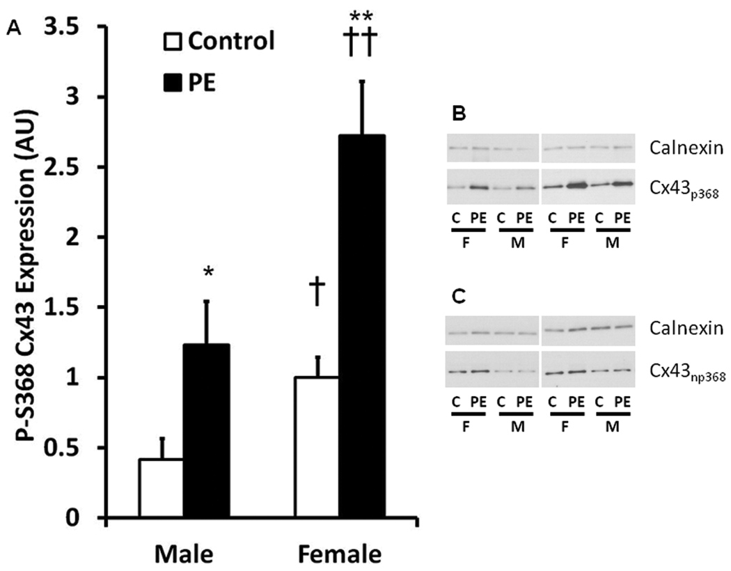
Decreases in cardiac connexin43 (Cx43) play a critical role in abnormal cell-to-cell communication and have been linked to the resistance of the female heart to arrhythmias. We therefore hypothesized that Cx43 expression would be greater in female cardiomyocytes than in male cardiomyocytes under pathologic conditions. Adult ventricular myocytes were isolated from male and female rats and treated with phenylephrine (PE), a well-established pathologic stimulus. Cx43 gene and protein expression was determined. The expression of micro-RNA-1 (miR-1), a micro-RNA known to control Cx43 protein expression in cardiomyocytes, was also determined. Cx43 mRNA and protein levels were significantly higher in the female cardiomyocytes than in the male cardiomyocytes (mRNA: 1.4-fold; Protein: 5-fold, both P < 0.05) under both basal and pathologic conditions. PE treatment increased Cx43 expression only in female cardiomyocytes. Cx43 phosphorylation, a marker of preserved Cx43 function, was also higher (P < 0.05), and The expression of miR-1 was lower (P < 0.05) in the female cardiomyocytes after PE treatment. The expression of miR-1 was unchanged by PE treatment in male cardiomyocytes. Thus, a sex difference in miR-1 may be responsible for the sex difference in Cx43 expression in cardiomyocytes under pathologic conditions. Taken together, our results demonstrate a sex difference in Cx43 expression and site-specific phosphorylation that favors cardioprotection in female cardiomyocytes.
Figures

Similar articles
-
MicroRNA-206 Downregulates Connexin43 in Cardiomyocytes to Induce Cardiac Arrhythmias in a Transgenic Mouse Model.Heart Lung Circ. 2019 Nov;28(11):1755-1761. doi: 10.1016/j.hlc.2018.09.008. Epub 2018 Oct 4. Heart Lung Circ. 2019. PMID: 30322759
-
Downregulation of connexin43 by microRNA-130a in cardiomyocytes results in cardiac arrhythmias.J Mol Cell Cardiol. 2014 Sep;74:53-63. doi: 10.1016/j.yjmcc.2014.04.024. Epub 2014 May 10. J Mol Cell Cardiol. 2014. PMID: 24819345 Free PMC article.
-
Signal transduction and transcriptional control of cardiac connexin43 up-regulation after alpha 1-adrenoceptor stimulation.J Pharmacol Exp Ther. 2008 Jul;326(1):315-22. doi: 10.1124/jpet.108.136663. Epub 2008 Apr 29. J Pharmacol Exp Ther. 2008. PMID: 18445782
-
Connexin 43 and Mitochondria in Cardiovascular Health and Disease.Adv Exp Med Biol. 2017;982:227-246. doi: 10.1007/978-3-319-55330-6_12. Adv Exp Med Biol. 2017. PMID: 28551790 Review.
-
[Remodeling of cardiac gap junctions and arrhythmias].Sheng Li Xue Bao. 2011 Dec 25;63(6):586-92. Sheng Li Xue Bao. 2011. PMID: 22193455 Review. Chinese.
Cited by
-
Omacor Protects Normotensive and Hypertensive Rats Exposed to Continuous Light from Increased Risk to Malignant Cardiac Arrhythmias.Mar Drugs. 2021 Nov 24;19(12):659. doi: 10.3390/md19120659. Mar Drugs. 2021. PMID: 34940658 Free PMC article.
-
Restoration of Adiponectin-Connexin43 Signaling Mitigates Myocardial Inflammation and Dysfunction in Diabetic Female Rats.J Cardiovasc Pharmacol. 2020 Mar;75(3):259-267. doi: 10.1097/FJC.0000000000000789. J Cardiovasc Pharmacol. 2020. PMID: 31868825 Free PMC article.
-
Hypertension Induces Pro-arrhythmic Cardiac Connexome Disorders: Protective Effects of Treatment.Biomolecules. 2023 Feb 9;13(2):330. doi: 10.3390/biom13020330. Biomolecules. 2023. PMID: 36830700 Free PMC article. Review.
-
Connexins in Cardiovascular and Neurovascular Health and Disease: Pharmacological Implications.Pharmacol Rev. 2017 Oct;69(4):396-478. doi: 10.1124/pr.115.012062. Pharmacol Rev. 2017. PMID: 28931622 Free PMC article. Review.
-
Cardiac myocyte alternans in intact heart: Influence of cell-cell coupling and β-adrenergic stimulation.J Mol Cell Cardiol. 2015 Jul;84:1-9. doi: 10.1016/j.yjmcc.2015.03.012. Epub 2015 Mar 28. J Mol Cell Cardiol. 2015. PMID: 25828762 Free PMC article.
References
-
- Akar FG, Nass RD, Hahn S, Cingolani E, Shah M, Hesketh GG, DiSilvestre D, Tunin RS, Kass DA, Tomaselli GF. Dynamic changes in conduction velocity and gap junction properties during development of pacing-induced heart failure. Am J Physiol Heart Circ Physiol. 2007;293:H1223–H1230. - PubMed
-
- Beardslee MA, Lerner DL, Tadros PN, Laing JG, Beyer EC, Yamada KA, Kleber AG, Schuessler RB, Saffitz JE. Dephosphorylation and intracellular redistribution of ventricular connexin43 during electrical uncoupling induced by ischemia. Circ Res. 2000;87:656–662. - PubMed
Publication types
MeSH terms
Substances
Grants and funding
LinkOut - more resources
Full Text Sources
