Review
. 2011 Jul 14;11(8):541-57.
doi: 10.1038/nrc3087.

Advances in sarcoma genomics and new therapeutic targets

Affiliations
Review

Advances in sarcoma genomics and new therapeutic targets

Barry S Taylor et al. Nat Rev Cancer. .

Erratum in

  • Nat Rev Cancer. 2011;11(9):685

Abstract

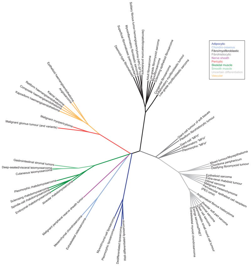
Increasingly, human mesenchymal malignancies are being classified by the abnormalities that drive their pathogenesis. Although many of these aberrations are highly prevalent within particular sarcoma subtypes, few are currently targeted therapeutically. Indeed, most subtypes of sarcoma are still treated with traditional therapeutic modalities, and in many cases sarcomas are resistant to adjuvant therapies. In this Review, we discuss the core molecular determinants of sarcomagenesis and emphasize the emerging genomic and functional genetic approaches that, coupled with novel therapeutic strategies, have the potential to transform the care of patients with sarcoma.


Conflict of interest statement

Competing interests statement

The authors declare no competing financial interests.

Figures

Figure 1
Figure 1. Taxonomy of soft tissue sarcoma
This unrooted phylogeny shows ~60 sarcoma subtypes as originally defined by the World Health Organization International Agency for Research on Cancer amended and updated based on current knowledge. The classification reflects relationships among lineage, prognosis (malignant, intermediate or locally aggressive, intermediate or rarely metastasizing), driver alterations, and additional parameters. Branch lengths determined by nearest neighbor joining of a discretized distance matrix based on aforementioned variables. Initial branching reflects differences in lineage with associated lineages appearing closer in distance (e.g. skeletal and smooth muscle). Subsequent branching encodes similarity in prognosis, whether they are translocation-associated, and if so, the genes shared among distinct fusions (in this order). While incomplete, as many subtypes lack sufficient global molecular profiling data on which to base a phylogeny, this initial formulation minimally reflects the relationships among lineage and major molecular lesions in the subtypes. The figure excludes 52 benign types of tumor. MFH, as abbreviated, represents undifferentiated pleomorphic sarcoma.
Figure 2
Figure 2. The structure of sarcoma genomes
A, Summary of recurrent translocations in malignant soft-tissue sarcomas indicates shared fusion partners between subtypes and regions of the genome subject to more frequent rearrangement. The outer ring represents genomic location (as labeled), and curves join fusion partners. B, Upper right: the somatic structure of an intermediate-genomic complexity sarcoma, a dedifferentiated liposarcoma (whole genome, inset) as defined by long-insert low depth-of-coverage mate-paired second-generation sequencing (see Box 1; Taylor BS and Singer S, unpublished data). Intra-chromosomal rearrangements are shown in gold and inter-chromosomal rearrangements in red; a subset of the interchromosomal rearrangements is reminiscent of rearranged sequence on chromosome 12 (chr12) in panel A. Lower left: the pathognomonic chromosome 12q amplification is shown in greater detail. This detailed view indicates a dense network of back-and-forth inverted and non-inverted intra-chromosomal rearrangements in three clusters (in grey, light blue, and dark blue; for clarity, inter-chromosomal rearrangements excluded). The curves reflect rearrangements between two genomic loci as determined experimentally and computationally. ACTB, actin-β; ALK, anaplastic lymphoma receptor tyrosine kinase; ASPSCR1, alveolar soft part sarcoma chromosome region candidate 1; ATF1, activating transcription factor 1; COL1A1, collagen type Iα1; CREB1, cAMP responsive element binding protein 1; CREB3L, AMP responsive element binding protein 3-like; DDIT3, DNA-damage-inducible transcript 3 (also known as CHOP); EPC1, enhancer of polycomb homolog 1; ETV6, ets variant 6; EWSR1, Ewing sarcoma breakpoint region 1; FLI1, Friend leukemia virus integration 1; FOXO1, forkhead box O1; FUS, fused in sarcoma; GLI1, GLI family zinc finger 1; JAZF1, JAZF zinc finger 1; NR4A3, nuclear receptor subfamily 4A3; NTRK3, neurotrophic tyrosine kinase receptor 3; PAX, paired box; PDGFB, platelet-derived growth factor-β; PHF1, PHD finger protein 1; SS18, synovial sarcoma translocation chromosome 18; SSX, synovial sarcoma, X breakpoint; SUZ12, suppressor of zeste 12; TAF15, TAF15 RNA polymerase II TATA box binding protein-associated factor; TCF12, transcription factor 12; TFE3, transcription factor binding to IGHM enhancer 3; TPM, tropomyosin; WT1, Wilms tumor 1.
Figure 3
Figure 3. Models and functional genetics
High-throughput integrative genomics, increasingly dominated by second-generation sequencing, identifies abnormalities of sequence, structure, expression, or copy number (A) in sarcoma genomes (notwithstanding epigenetic modification). In parallel, model systems (cultured cells, animal models, or tissue slices) need to be generated from human tumors (B) and similarly genomically profiled to confirm that they represent primary tumors in the retention of driver alterations (C). From tumor profiles, computational methodologies analyze the patterns of recurrence to distinguish likely driver from passenger alterations (D) (Box 2). These models can then be subjected to high-throughput functional genetic analyses, including both gain-of-function approaches (such as open reading frame (ORF) overexpression with pLX-Blast-V5 or similar cDNA expression vectors) and loss-of-function approaches (such as RNA interference using pLKO1-puro or similar shRNA plasmids) (E). These approaches can be applied either to all genes (to identify genotype-selective targets from those that are not) or to those identified by the integrative genomic and statistical methods. The readouts of these high throughput methods, be it fluorescence, barcode arrays, or sequence read counts, provide data on one of a large number of possible phenotypes and can be analyzed (F) to identify genotype-dependent vulnerabilities in sarcoma cells, identifying targets that can be validated in orthogonal models (G), a subset of which may be suitable for therapeutic intervention. This model of genomics-driven functional genetics focuses on rapid functional annotation of cancer genomes. GEMM, genetically engineered mouse model.
Figure 4
Figure 4. Pathways for targeted therapy in sarcoma
Diverse subtype-specific alterations imply that a variety of signaling pathways function aberrantly in sarcomas. Abbreviated pathways include Ras-Raf, PI3K, mTOR, p53, cell cycle and survival, Notch, and Hedgehog signaling, all of which are targeted by a growing list of specific therapies. Here, annotation of nodes in signaling networks affected by specific genomic abnormalities includes affected subtype, alteration types (genomic amplification or deletion are solid triangles, over- or under-expressed are open arrowheads, mutated are starred), and frequencies. A subset of nodes are colored by their dominant alteration type (see key). Targeted agents (gray) include those in clinical use and those in preclinical or early-phase development in sarcoma. CCND, cyclin D; CDK, cyclin-dependent kinase; CSL, recombination signal binding protein for immunoglobulin kappa J region (also known as RBPJ); DDLPS, dedifferentiated liposarcoma; EGFR, epidermal growth factor receptor; GIST, gastrointestinal stromal tumor; HDAC, histone deacetylase; HPC-SFT, hemangiopericytoma-solitary fibrous tumor; HSP90, heat shock protein 90; IGF1R, insulin-like growth factor 1 receptor; NICD, NOTCH intracellular domain; NF1, neurofibromin 1; PDGFR, platelet-derived growth factor receptor; PEComa, perivascular epitheliod cell tumor; PTCH, patched; RB1, retinoblastoma 1; RHEB, Ras homolog enriched in brain; SMO, smoothened; SSH, slingshot; TSC2, tuberin; VEGFR2, vascular endothelial growth factor receptor 2; WDLPS, well-differentiated liposarcoma.

References

    1. Fletcher C, Unni K, Mertens F. Pathology and genetics of tumors of soft tissue and bone. International Agency for Research on Cancer Press; Lyon: 2002.
    1. Jemal A, Siegel R, Xu J, Ward E. Cancer statistics, 2010. CA Cancer J Clin. 2010;60:277–300. - PubMed
    1. Borden EC, et al. Soft tissue sarcomas of adults: state of the translational science. Clin Cancer Res. 2003;9:1941–56. - PubMed
    1. Helman LJ, Meltzer P. Mechanisms of sarcoma development. Nat Rev Cancer. 2003;3:685–94. - PubMed
    1. Mertens F, et al. Translocation-related sarcomas. Semin Oncol. 2009;36:312–23. - PubMed

Publication types

Substances