. 2012:2012:518426.
doi: 10.1155/2012/518426. Epub 2011 Jun 28.

A PEDF-derived peptide inhibits retinal neovascularization and blocks mobilization of bone marrow-derived endothelial progenitor cells

Affiliations

A PEDF-derived peptide inhibits retinal neovascularization and blocks mobilization of bone marrow-derived endothelial progenitor cells

Richard Longeras et al. Exp Diabetes Res. 2012.

Abstract

Proliferative diabetic retinopathy is characterized by pathological retinal neovascularization, mediated by both angiogenesis (involving mature endothelial cells) and vasculogenesis (involving bone marrow-derived circulating endothelial progenitor cells (EPCs)). Pigment epithelium-derived factor (PEDF) contains an N-terminal 34-amino acid peptide (PEDF-34) that has antiangiogenic properties. Herein, we present a novel finding that PEDF-34 also possesses antivasculogenic activity. In the oxygen-induced retinopathy (OIR) model using transgenic mice that have Tie2 promoter-driven GFP expression, we quantified Tie2GFP(+) cells in bone marrow and peripheral blood by fluorescence-activated cell sorting (FACS). OIR significantly increased the number of circulating Tie2-GFP(+) at P16, correlating with the peak progression of neovascularization. Daily intraperitoneal injections of PEDF-34 into OIR mice decreased the number of Tie2-GFP(+) cells in the circulation at P16 by 65% but did not affect the number of Tie2-GFP(+) cells in the bone marrow. These studies suggest that PEDF-34 attenuates EPC mobilization from the bone marrow into the blood circulation during retinal neovascularization.


Figures
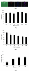
Figure 1
Figure 1
PEDF-34 acts in a concentration-dependent manner to inhibit cell proliferation and induce apoptosis specifically in endothelial cells. (a) The purity of primary BRCEC was examined using Dil-Ac-LDL uptake assays. The cells were counter-stained with DAPI, and exhibited a >99% purity based on positive Dil-Ac-LDL uptake. Both rat Müller cells (b) and BRCEC (c) were treated with increasing concentrations of PEDF 34-mer ranging from 25 to 400 nM. Viable cells were quantified after 72 h by MTT assay and expressed as % of the cells in control treated with the BSA only (denoted as NT on graph). (d) BRCECs were treated with increasing concentrations of PEDF 34-mer for 24 h. Apoptotic cells were quantified by counting Annexin V positive cells using FACS and expressed as % in total cells (mean + SD, n = 3).
Figure 2
Figure 2
Antiangiogenic activity of PEDF 34-mer during in vivo blood vessel formation. CAMs were separately treated with either 50 or 100 nM PEDF-34. (a) Left: close-up image of a chicken embryo CAM with a nitrocellulose disk containing PEDF-34 (100 nM). Middle: the disk area after removal of PEDF-34 nitrocellulose disk. Right: image of a PBS-treated-nitrocellulose control area on the same CAM with the disk removed. Note considerable reduction in the overall number of small newly formed blood vessels, when compared with the PBS-treated control. (b) Blood vessel density in the CAMs was assessed by software analysis. Vascular densities in the disk areas of CAMs treated with 50 and 100 nM were quantified and averaged. The graph represents the percent of vascular density found under PEDF-34-treated areas compared to the PBS-treated control area, which was set at 100%. Vascular density in the CAM treated with PEDF-34 is significantly lower than the control (mean + SD, n = 5).
Figure 3
Figure 3
Systemic injection of PEDF-34 reduced ischemia-induced retinal neovascularization. The graph represents the average number of preretinal (vitreous) vascular cells in OIR mice treated with BSA or PEDF-34. (mean ± SEM, BSA n = 4. PEDF-34 n = 5).
Figure 4
Figure 4
Isolation of Tie2-GFP+ cells from bone marrow and peripheral blood in mice. (a) Isolation of bone marrow and peripheral blood cells was performed in parallel as depicted. Washed cells were resuspended in ice-cold PBS and shielded from the light to prevent photobleaching. (b) and (c) Flow cytometric analysis of isolated cells. Resuspended cells were subjected to FACS on an Influx cell sorter. The profile obtained from Tie2-GFP mice (c) exhibited an additional population when compared to the wild-type C57BL/6 (b) (denoted in the shaded pink box). Therefore, this exclusive population was characteristic of the Tie2-GFP+ cells. (d) Tie2-GFP and Tie2-GFP+ cells as identified by FACS analysis were collected separately, fixed onto slides, counter-stained with DAPI, and visualized by fluorescence microscopy. Only cells identified by prior FACS analysis as Tie2-GFP+ exhibited significant levels of green fluorescence by microscopic (100x) analysis. (e) Tie2-GFP+ cells which were isolated by FACS from Tie2-GFP mice were viable and of the endothelial cell lineage, as demonstrated by their ability to uptake Dil-Ac-LDL. Cells sorted by FACS as Tie2-GFP did not have the ability to uptake Dil-Ac-LDL.
Figure 5
Figure 5
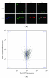
Characterization of circulating Tie2-GFP+ cells as EPCs. (a) Tie2-GFP+ cells from Tie2-GFP mice were sorted onto slides and immunostained for the EPC-specific markers CD117 and CD133 and counterstained with DAPI. Confocal microscopy was used to capture images at 40x magnification; (b) EPCs were fixed, sorted for GFP, then immunostained for CD117, and subjected to FACS. Most Tie2-GFP+ cells stained positive for CD117, indicating the majority of circulating endothelial cells are EPCs.
Figure 6
Figure 6
The number of circulating endothelial cells and EPCs increases in OIR mice at P16. Tie2-GFP+ cells in both the bone marrow (BM) and peripheral blood (PB) from Tie2-GFP mice were quantified by FACS at p12, p16 and p20 under normoxic rearing conditions (grey), or in the OIR model (black). Graphs represent the percent of Tie2-GFP+ cells based on 100% being set to the average number of Tie2-GFP+ cells from normoxic mice.
Figure 7
Figure 7
PEDF-34 reduces the number of circulating endothelial cells and EPCs in OIR mice. In the OIR model, Tie2-GFP pups received a daily i.p. injection of either PEDF-34, BSA, or a control peptide in PBS at a dose of 5 mg/kg of body weight from P12 to P16. At P16, Tie2-GFP+ circulating endothelial cells and EPCs in peripheral blood were quantified by FACS. The graph represents the percentage of Tie2-GFP+ cells in peripheral blood, based on 100% being set to the average number of Tie2-GFP+ cells from OIR mice injected with BSA alone.

Similar articles

Cited by

References

    1. Battegay EJ. Angiogenesis: mechanistic insights, neovascular diseases, and therapeutic prospects. Journal of Molecular Medicine. 1995;73(7):333–346. - PubMed
    1. Bouck N. PEDF: anti-angiogenic guardian of ocular function. Trends in Molecular Medicine. 2002;8(7):330–334. - PubMed
    1. Gao G, Li Y, Zhang D, Gee S, Crosson C, Ma JX. Unbalanced expression of VEGF and PEDF in ischemia-induced retinal neovascularization. FEBS Letters. 2001;489(2-3):270–276. - PubMed
    1. Miller JW, Adamis AP, Aiello LP. Vascular endothelial growth factor in ocular neovascularization and proliferative diabetic retinopathy. Diabetes Metabolism Reviews. 1997;13(1):37–50. - PubMed
    1. Abu El-Asrar AM, Struyf S, Verbeke H, Van Damme J, Geboes K. Circulating bone-marrow-derived endothelial precursor cells contribute to neovascularization in diabetic epiretinal membranes. Acta Ophthalmologica. 2011;89(3):222–228. - PubMed

Publication types

MeSH terms