

Pneumomediastinum as first manifestation of emphysematous pyelonephritis in a patient who is non-diabetic
- PMID: 21754958
- PMCID: PMC3028289
- DOI: 10.1136/bcr.05.2009.1873
Pneumomediastinum as first manifestation of emphysematous pyelonephritis in a patient who is non-diabetic
Abstract
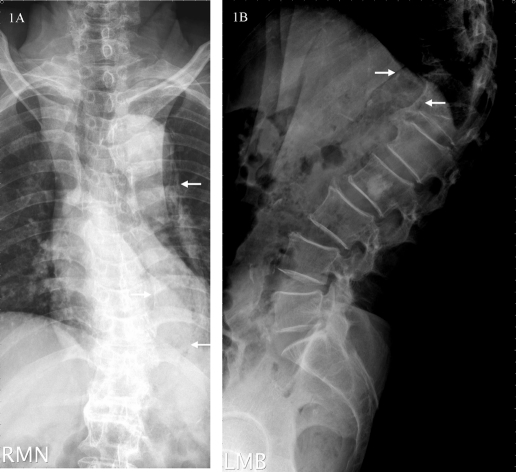
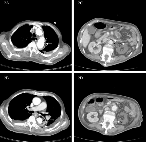
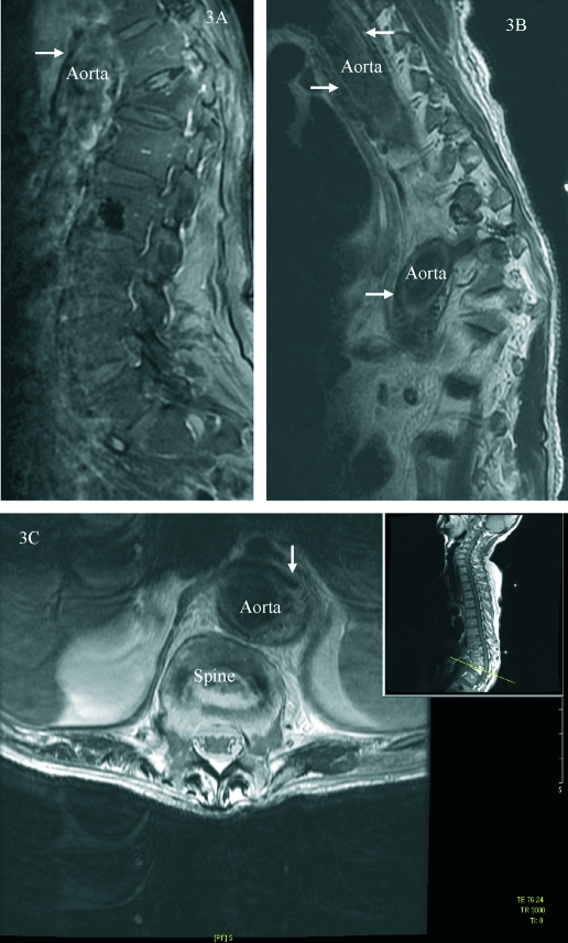
Emphysematous pyelonephritis (EPN) is a rare but life-threatening acute suppurative infection of the kidney, characterised by production of gas within the renal parenchyma, collecting system or perirenal tissue. It has a high mortality rate (70% to 90%), and the majority of patients have diabetes mellitus. The left kidney is most common involved and Escherichia coli is the most common pathogen. EPN complicated with pneumomediastinum (PM) has been reported in only four cases previously. Here, a case of PM as first manifestation of EPN in a non-diabetic 81-year-old man is reported. He had experienced back pain and abdominal fullness for 1 week. A plain radiograph, CT aortography and MRI confirmed the diagnosis of EPN complicated with PM. The patient died on the 22nd day of treatment with antibiotics of cefmetazole, gentamycin and metronidazole.
Figures
References
-
- Ting KH, Lin KH, Chang CC. Emphysematous pyelonephritis: presenting with pneumomediastinum. Am J Emerg Med 2006; 24: 350–2 - PubMed
-
- Park SH, Hong HP, Kim MC, et al. Emphysematous pyelonephritis associated with pneumoperitoneum and pneumomediastinum: a case report. J Korean Soc Emerg Med 2005; 16: 398–402
-
- Wang YC, Wang JM, Chow YC, et al. Pneumomediastinum and subcutaneous emphysema as the manifestation of emphysematous pyelonephritis. Int J Urol 2004; 11: 909–11 - PubMed
-
- Menif E, Nouira K, Baccar S, et al. Emphysematous pyelonephritis: report of 3 cases. Ann Urol (Paris) 2001; 35: 97–100 - PubMed
-
- Huang JJ, Tseng CC. Emphysematous pyelonephritis: clinicoradiological classification, management, prognosis, and pathogenesis. Arch Intern Med 2000; 160: 797–805 - PubMed
LinkOut - more resources
Full Text Sources