. 2011;6(7):e21774.
doi: 10.1371/journal.pone.0021774. Epub 2011 Jul 6.

Vascular endothelial expression of indoleamine 2,3-dioxygenase 1 forms a positive gradient towards the feto-maternal interface

Affiliations

Vascular endothelial expression of indoleamine 2,3-dioxygenase 1 forms a positive gradient towards the feto-maternal interface

Astrid Blaschitz et al. PLoS One. 2011.

Abstract

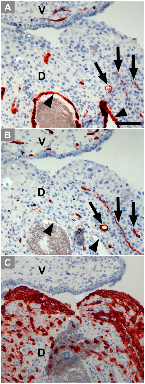
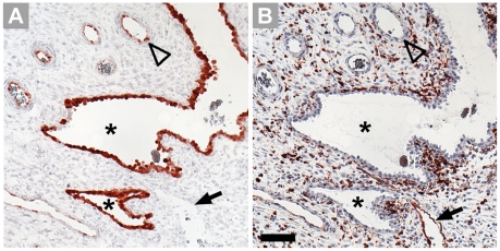
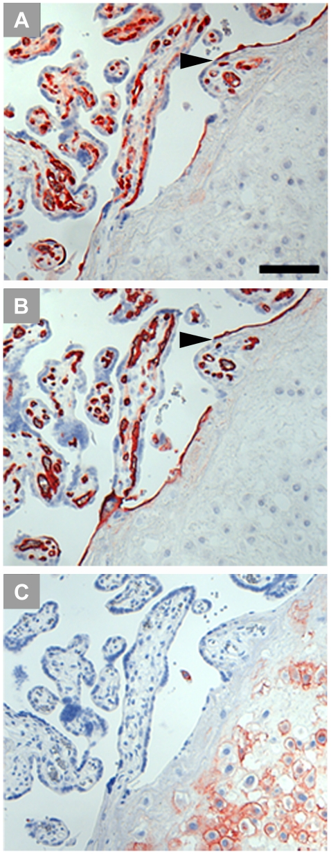
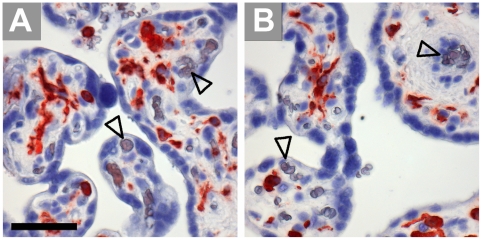
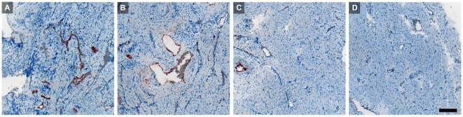
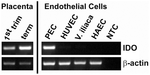
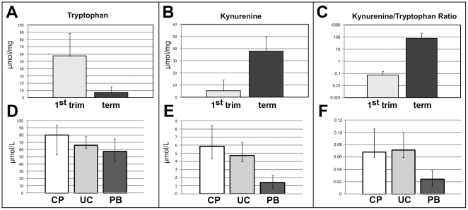
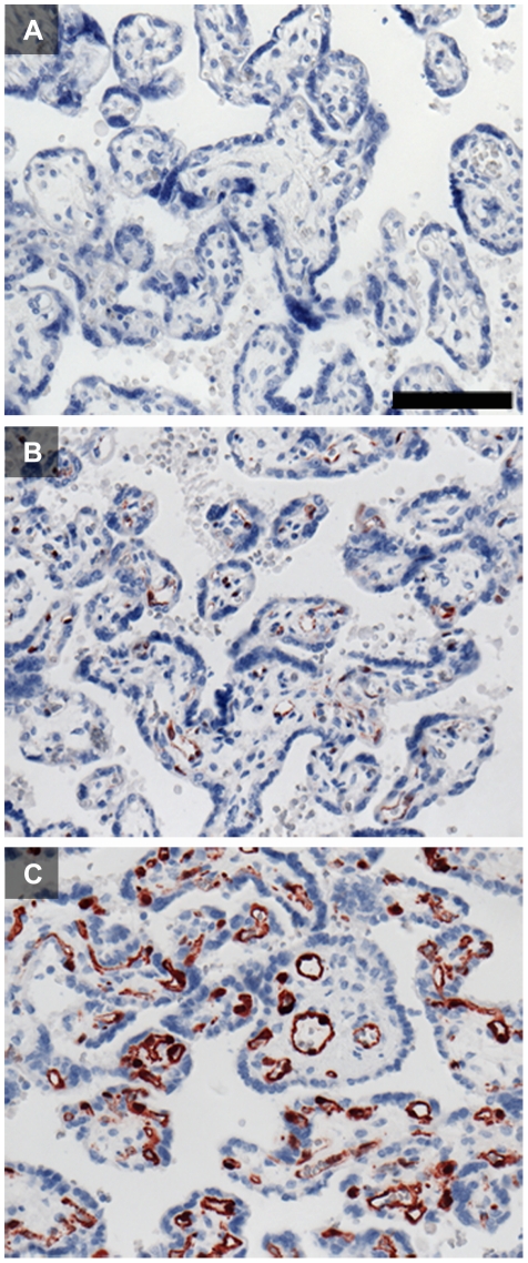
We describe the distribution of indoleamine 2,3-dioxygenase 1 (IDO1) in vascular endothelium of human first-trimester and term placenta. Expression of IDO1 protein on the fetal side of the interface extended from almost exclusively sub-trophoblastic capillaries in first-trimester placenta to a nearly general presence on villous vascular endothelia at term, including also most bigger vessels such as villous arteries and veins of stem villi and vessels of the chorionic plate. Umbilical cord vessels were generally negative for IDO1 protein. In the fetal part of the placenta positivity for IDO1 was restricted to vascular endothelium, which did not co-express HLA-DR. This finding paralleled detectability of IDO1 mRNA in first trimester and term tissue and a high increase in the kynurenine to tryptophan ratio in chorionic villous tissue from first trimester to term placenta. Endothelial cells isolated from the chorionic plate of term placenta expressed IDO1 mRNA in contrast to endothelial cells originating from human umbilical vein, iliac vein or aorta. In first trimester decidua we found endothelium of arteries rather than veins expressing IDO1, which was complementory to expression of HLA-DR. An estimation of IDO activity on the basis of the ratio of kynurenine and tryptophan in blood taken from vessels of the chorionic plate of term placenta indicated far higher values than those found in the peripheral blood of adults. Thus, a gradient of vascular endothelial IDO1 expression is present at both sides of the feto-maternal interface.


Conflict of interest statement

Competing Interests: The authors have declared that no competing interests exist.

Figures

Figure 1
Figure 1. Immunohistochemical staining of first trimester placenta serial paraffin sections at the 11th week of pregnancy.
Villous chorion (A, B) and umbilical cord vessels (C, D) stained for IDO1 (A, C) and for the endothelial marker CD34 (B, D); bigger vessels located more to the center of the villi are indicated by arrowheads, capillaries forming a network beneath the trophoblastic double layer stain positive for IDO1. The scale bar represents 100 µm.
Figure 2
Figure 2. Immunohistochemical staining of serial paraffin sections of first trimester decidua basalis (D) showing also partly villous tissue (V).
The sections are stained for IDO1 (A), the endothelial marker CD34 (B) and HLA–G, a marker for extravillous trophoblast (C). Arrow heads heads point towards the epithelium of uterine glands, some endothelia are indicated by arrows. The scale bar represents 100 µm.
Figure 3
Figure 3. Comparison of localization of IDO1 (A) and HLA-DR (B) in first trimester decidua.
Serial paraffin sections are stained by immunohistochemistry. Designated are uterine glands (asterisks), the endothelium of spiral arteries (open arrowhead) and of veins (arrow). Please note that apart from expression in the endothelium of veins, HLA-DR is expressed by macrophages. Scale bar = 100 µm.
Figure 4
Figure 4. Immunohistochemical staining of term placenta serial sections including the basal plate.
IDO1 labelling results in staining of vascular endothelium, leaving villous and extravillous trophoblast unstained (A). The anti-CD34 antibody stains endothelial cells of the fetal circulation in the villous mesenchymal core and also endothelial cells which line the intervillous space; the arrowhead indicates a special site where endothelium connects with syncytiotrophoblast (B). HLA-G identifies extravillous cytotrophoblasts located in the basal plate (C). Scale bar represents 100 µm.
Figure 5
Figure 5. Immunohistochemical staining of paraffin sections of term villous tissue.
Expression of HLA-DR (A) is allocated to Hofbauer cells, as demonstrated in a serial section stained for the macrophage marker CD163 (B), open arrowheads indicate vascular endothelia. Scale bars = 50 µm.
Figure 6
Figure 6. Immunohistochemical staining of serial paraffin sections of term placenta chorionic plate (A, B) and umbilical cord (C, D).
The sections are stained for IDO1 (A, C) and the endothelial cell marker CD34 (B, D).Scale bar represents 100 µm.
Figure 7
Figure 7. Paraffin sections of a uterine wall including the decidua in the 22nd week of gestation, stained with anti-IDO1 antibody.
The trophoblast-invaded implantation site shows strong staining of vascular endothelial cells (A). In the second quarter of the uterine wall most of the myometrial blood vessels are also labeled (B). The third quarter shows only one vessel stained (C) and in the outermost quarter of the uterus (D) none of the vessels stain for IDO1. Scale bar represents 200 µm.
Figure 8
Figure 8. Expression of IDO1 mRNA.
Semi-quantitative RT-PCR comparing IDO1 mRNA expression in first trimester and term placenta tissues with endothelial cells isolated from term placenta chorionic plate (PEC), a human umbilical vein endothelial cell line (HUVEC), endothelial cells isolated from the iliac vein (V. iliaca) and a cell line from human aortic endothelium (HAEC). The expression of beta-actin was used as internal reference and water instead of total RNA served as non template control (NTC).
Figure 9
Figure 9. Chorionic tryptophan-degrading activity.
Concentration of tryptophan (A, D), kynurenine (B, E) and the kyn/trp ratio (C, F) in placenta villous tissues (A, B, C) at first trimester (grey bars) and at term (black bars; indicated are the means+SD. For all three comparisons p<0.001) and in blood samples (D, E, F) from chorionic plate vessels of term placenta (CP, white bars), from umbilical cord blood (UC, light grey bars) and from peripheral blood of healthy blood donors (PB, dark grey bars). D–F: Indicated are the medians and the ranges. Assuming a significance level of 0.01, the test results for all three parameters are significant. The difference between the groups CP and PB is significant for all three parameters at p<0.001.
Figure 10
Figure 10. Illustration of the IDO1 expression gradient in vascular endothelial cells of fetal and maternal blood circulations at the utero-placental materno-fetal interface.
The expression intensity increases close to the semi-allogeneic contact zone.
Figure 11
Figure 11. Influence of heat induced epitope retrieval (HIER) on IDO1 staining of formalin-fixed paraffin-embedded term placenta sections.
Staining without any pre-treatment (A) does not allow for detection of IDO1. IDO1 can be localized in endothelial cells of the villous chorion after HIER using citrate buffer at pH 6 (B). HIER with retrieval buffer at pH 9 (C) enhanced the staining intensity and allows for a more detailed interpretation of IDO1 immunolocalization. The scale bar represents 100 µm.

References

    1. Munn DH, Shafizadeh E, Attwood JT, Bondarev I, Pashine A, et al. Inhibition of T cell proliferation by macrophage tryptophan catabolism. J Exp Med. 1999;189:1363–1372. - PMC - PubMed
    1. Munn DH, Zhou M, Attwood JT, Bondarev I, Conway SJ, et al. Prevention of allogeneic fetal rejection by tryptophan catabolism [see comments]. Science. 1998;281:1191–1193. - PubMed
    1. Koch CA, Platt JL. T cell recognition and immunity in the fetus and mother. Cell Immunol. 2007;248:12–17. - PMC - PubMed
    1. Hönig A, Rieger L, Kapp M, Sutterlin M, Dietl J, et al. Indoleamine 2,3-dioxygenase (IDO) expression in invasive extravillous trophoblast supports role of the enzyme for materno-fetal tolerance. J Reprod Immunol. 2004;61:79–86. - PubMed
    1. Kudo Y, Boyd CA, Spyropoulou I, Redman CW, Takikawa O, et al. Indoleamine 2,3-dioxygenase: distribution and function in the developing human placenta. J Reprod Immunol. 2004;61:87–98. - PubMed

Publication types

MeSH terms