. 2011;6(7):e21930.
doi: 10.1371/journal.pone.0021930. Epub 2011 Jul 6.

Small compound 6-O-angeloylplenolin induces mitotic arrest and exhibits therapeutic potentials in multiple myeloma

Affiliations

Small compound 6-O-angeloylplenolin induces mitotic arrest and exhibits therapeutic potentials in multiple myeloma

Ying Liu et al. PLoS One. 2011.

Abstract

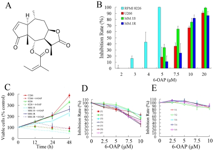
Background: Multiple myeloma (MM) is a disease of cell cycle dysregulation while cell cycle modulation can be a target for MM therapy. In this study we investigated the effects and mechanisms of action of a sesquiterpene lactone 6-O-angeloylplenolin (6-OAP) on MM cells.

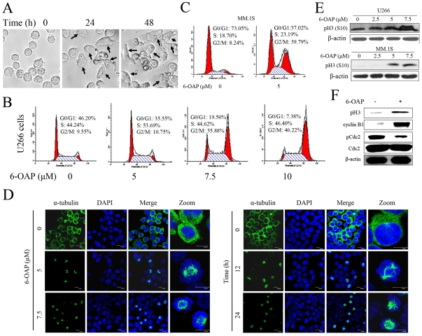
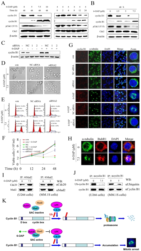
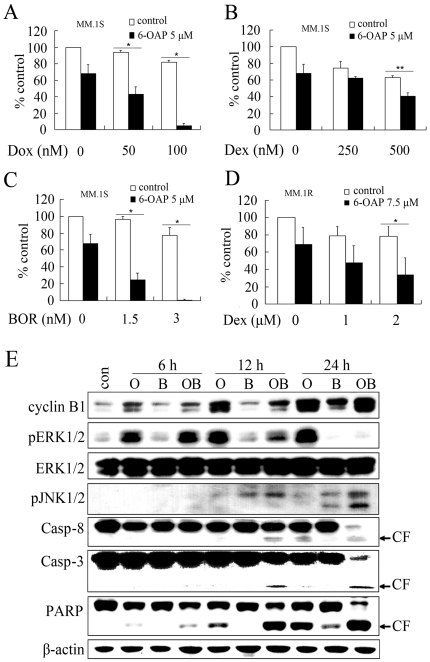
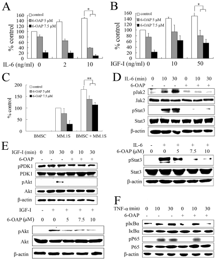
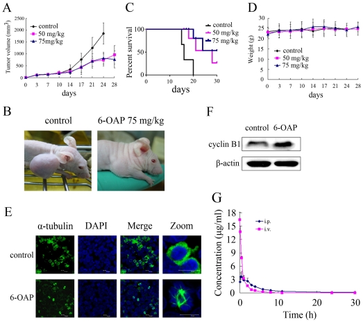
Methodology/principal findings: MM cells were exposed to 6-OAP and cell cycle distribution were analyzed. The role for cyclin B1 to play in 6-OAP-caused mitotic arrest was tested by specific siRNA analyses in U266 cells. MM.1S cells co-incubated with interleukin-6 (IL-6), insulin-like growth factor-I (IGF-I), or bone marrow stromal cells (BMSCs) were treated with 6-OAP. The effects of 6-OAP plus other drugs on MM.1S cells were evaluated. The in vivo therapeutic efficacy and pharmacokinetic features of 6-OAP were tested in nude mice bearing U266 cells and Sprague-Dawley rats, respectively. We found that 6-OAP suppressed the proliferation of dexamethasone-sensitive and dexamethasone-resistant cell lines and primary CD138+ MM cells. 6-OAP caused mitotic arrest, accompanied by activation of spindle assembly checkpoint and blockage of ubiquitiniation and subsequent proteasomal degradation of cyclin B1. Combined use of 6-OAP and bortezomib induced potentiated cytotoxicity with inactivation of ERK1/2 and activation of JNK1/2 and Casp-8/-3. 6-OAP overcame the protective effects of IL-6 and IGF-I on MM cells through inhibition of Jak2/Stat3 and Akt, respectively. 6-OAP inhibited BMSCs-facilitated MM cell expansion and TNF-α-induced NF-κB signal. Moreover, 6-OAP exhibited potent anti-MM activity in nude mice and favorable pharmacokinetics in rats.

Conclusions/significance: These results indicate that 6-OAP is a new cell cycle inhibitor which shows therapeutic potentials for MM.


Conflict of interest statement

Competing Interests: The authors have declared that no competing interests exist.

Figures

Figure 1
Figure 1. 6-OAP inhibits cell proliferation of MM cells.
(A) Chemical structure of 6-OAP. (B) Effects of 6-OAP on cell proliferation of RPMI 8226, U266, MM.1S, and MM.1R cells. In this experiment, cells were treated with 6-OAP at indicated concentration for 48 h, and analyzed by MTT assay. (C) MM cells were treated with or without 6-OAP, and analyzed by trypan blue exclusion assay. P values for difference between the cells treated without and with 6-OAP for 48 h: p<.0001 for U266 and RPMI 8226 cells; p = .0037 for MM.1S and p = .0043 for MM.1R cells. (D) CD138+ cells harvested from 7 MM patients (P1–P7) were treated with or without 6-OAP, and analyzed by MTT assay. (E) Peripheral blood mononuclear cells from 5 healthy volunteers (V1-V5) were co-incubated with 6-OAP at indicated concentration for 48 h, and analyzed by MTT assay.
Figure 2
Figure 2. 6-OAP induces mitotic arrest in MM cells.
(A) U266 cells were treated with 6-OAP at 7.5 µM for 0, 24, 48 h and imaged by Leica DMI 400B microscope. (B) U266 cells were treated with 6-OAP at indicated concentrations for 48 h. Cell cycle distribution was determined by flow cytometry. (C) MM.1S cells were treated with 5 µM 6-OAP for 48 h. Cell cycle distribution was determined by flow cytometry. (D) U266 cells were treated with 6-OAP at 5 to 7.5 µM for 24 h (left panel) or at 7.5 µM for 12 to 24 h (right panel). For immunofluorescence analysis of microtubules, cells was stained with an anti-α-tubulin antibody to visualize microtubules (green) and DAPI to counter stained DNA (blue) and observed by confocal microscopy. (E) Indicated cells were treated with 6-OAP at indicated concentration for 24 h, lysed, and Western blotting was performed using antibodies indicated. (F) CD138+ primary cells isolated from 1 MM patient were treated with 6-OAP (7.5 µM) at indicated concentration for 24 h, lysed, and Western blotting was performed using antibodies indicated.
Figure 3
Figure 3. 6-OAP accumulates cyclin B1 which is required for mitotic arrest.
(A) U266 cells were treated with 6-OAP at indicated concentration and time points, lyzed, and Western blotting was performed using antibodies indicated. (B) MM.1S cells were treated with 6-OAP for 48 h, lyzed, and Western blotting was conducted using antibodies indicated. (C) U266 cells were transfected with 50 nM cyclin B1-specific siRNA1, siRNA2 or NC siRNA, followed by treatment without or with 6-OAP. Cells were harvested for Western blot analyses. (D through F) U266 cells were transfected with cyclin B1-specific siRNA2 or NC siRNA, followed by treatment with 6-OAP at 7.5 µM. After co-incubation with 6-OAP for 18 h, the cells were then harvested and detected by Leica DMI 400B microscope (D), or analyzed by flow cytometry for the cell cycle distribution (E). To evaluate cell growth, the cells were treated with 6-OAP for indicated time points and analyzed by trypan blue exclusion assay (F). (G) U266 cells were transfected with cyclin B1-specific siRNA1 or NC siRNA, followed by treatment with 6-OAP at 7.5 µM for 18 h. The cells were assayed by immunofluorescence analysis using an anti-cyclin B1 antibody to visualize the expression of cyclin B1 (red), an anti-α-tubulin antibody to visualize microtubules (green), and DAPI to counter stained DNA (blue). (H) U266 cells were treated without or with 7.5 µM 6-OAP for 24 h, and stained with anti-α-tubulin and anti-BubR1 antibodies to visualize microtubules (green), BubR1 (red), and DAPI to counter stained DNA (blue), and analyzed by confocal microscopy. (I and J) U266 and MM.1S cells were incubated without or with 6-OAP at indicated concentration for 24 h, lysed, and immunoprecipitation was performed followed by Western blotting using indicated antibodies. (K) Hypothetical model showing how 6-OAP causes cyclin B1 accumulation in MM cells. In eukaryocytes, APC can attach monoubiquitin to multiple lysine residues on cyclin B1, followed by polyubiquitin chain extensions linked through multiple lysine residues of ubiquitin . In this simplified model only one polyubiquitin chain is shown. Ub, ubiquitin.
Figure 4
Figure 4. 6-OAP enhances cytotoxicity of Dox, Dex and BOR to MM cells.
(A through C) MM.1S cells were treated for 48 h with Dox (A), Dex (B) or BOR (C) in the presence or absence of 6-OAP at 5 µM. MTT assay was used to test the proliferation of MM.1S cells. (D) MM.1R cells were treated for 48 h with Dex in the presence or absence of 6-OAP at 7.5 µM. MTT assay was used to test the proliferation of MM.1R cells. *p<.001,** p<.05. (E) MM.1S cells were cultured with control media (con), 6-OAP (O, 5 µM), BOR (B, 3 nM), or 6-OAP (5 µM) plus BOR (3 nM) (OB) for indicated time points. Cells were then lysed and subjected to Western blotting using indicated antibodies. CF, cleavage fragment.
Figure 5
Figure 5. 6-OAP overcomes the protective effects of IL-6, IGF-I and BMSCs on MM cells.
(A and B) MM.1S cells were cultured for 48 h with indicated concentrations of 6-OAP at the presence or absence of IL-6 (A) or IGF-I (B). Cell proliferation was assessed by MTT assay. (C) MM.1S and/or BMSCs cells were cultured for 48 h with indicated concentrations of 6-OAP. Cell proliferation was assessed by MTT assay. *p<.01,** p<.05. (D) MM.1S cells were serum starved for 2 h, then co-cultured without or with 6-OAP at 5 µM for 12 h, followed by stimulation with IL-6 at 10 ng/ml for indicated time points (upper panel), or IL-6 at 10 ng/ml for 1 h, followed by treatment with 6-OAP at indicated concentration for 12 h (lower panel). Whole-cell extracts were prepared and examined by Western blotting using antibodies against pJak2, pStat3, Stat3 or β-actin. (E) MM.1S cells were serum starved for 2 h, then co-cultured without or with 6-OAP (5 µM) for 12 h, followed by stimulation with IGF-I at 50 ng/ml for indicated time points (upper panel), or stimulated with IGF-I at 50 ng/ml for 1 h, followed by treatment with 6-OAP at indicated concentration for 12 h (lower panel). Whole-cell extracts were prepared and examined by western blotting using antibodies against pPDK1, pAkt, Akt or β-actin. (F) MM.1S cells were serum starved for 2 h, then co-cultured without or with 6-OAP (5 µM) for 12 h, followed by stimulation with TNF-α at 5 ng/ml for indicated time points. Whole-cell extracts were prepared and examined by Western blotting using indicated antibodies.
Figure 6
Figure 6. In vivo therapeutic efficacy of 6-OAP on human MM murine model.
Nude mice were given subcutaneous inoculations in the right flank with 1×10⁷ U266 cells. When the tumors reached a palpable size, the mice were treated intraperitoneally with vehicle or 6-OAP (50 or 75 mg/kg, 5 times a week for 4 weeks). (A) 6-OAP significantly inhibited MM tumor growth (P<.0001, 50 or 75 mg/kg vs control). (B) Growth inhibition of subcutaneous tumors was observed in mice treated with 6-OAP. (C) Survival curve of control and 6-OAP-treated mice. (D) Treatment with 6-OAP did not affect animal body weight. (E) Tumor samples were harvested from mice treated with vehicle or 50 mg/kg 6-OAP and subjected to immunofluorescence analysis using an anti-α-tubulin antibody and DAPI. (F) Tumor tissues were harvested from mice treated with vehicle or 50 mg/kg 6-OAP, whole-tissue lysates were subjected to Western blotting using an anti-cyclin B1 antibody. (G) The concentration-time profiles of 6-OAP after intravenous (30 mg/kg) or intraperitoneal (40 mg/kg) injection of 6-OAP in Sprague-Dawley rats.

References

    1. Kyle RA, Rajkumar SV. Multiple Myeloma. N Engl J Med. 2004;351:1860–1873. - PubMed
    1. Raab MS, Podar K, Breitkreutz I, Richardson PG, Anderson KC. Multiple myeloma. Lancet. 2009;374:324–339. - PubMed
    1. Parkin DM, Bray F, Ferlay J, Pisani P. Global cancer statistics, 2002. CA Cancer J Clin. 2005;55:74–108. - PubMed
    1. Dimopoulos M, Spencer A, Attal M, Prince HM, Harousseau JL, et al. Lenalidomide plus dexamethasone for relapsed or refractory multiple myeloma. N Engl J Med. 2007;357:2123–2132. - PubMed
    1. Richardson PG, Barlogie B, Berenson J, Singhal S, Jagannath S, et al. A phase 2 study of bortezomib in relapsed, refractory myeloma. N Engl J Med. 2003;348:2609–2617. - PubMed

Publication types

MeSH terms