

Symptomatic spinal cavernous malformations: indication for microsurgical treatment and outcome
- PMID: 21755413
- PMCID: PMC3175884
- DOI: 10.1007/s00586-011-1898-z
Symptomatic spinal cavernous malformations: indication for microsurgical treatment and outcome
Abstract
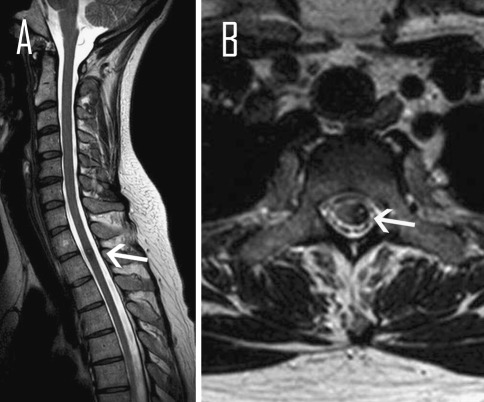
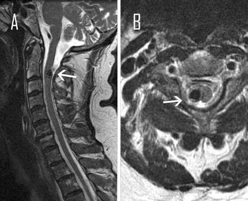
Purpose: We demonstrate clinical features, therapy and outcome of 14 patients with symptomatic spinal cavernous malformations (CM).
Methods: We retrospectively reviewed all patients who underwent microsurgical treatment of symptomatic spinal CM during the last decade in our department through an analysis of our database.
Results: We analyzed the data of 14 patients (11 females, 3 males) with symptomatic spinal CM in a range of 16-77 years (mean age 47.8 years). Seven patients (50%) experienced significant improvement of their symptoms rapidly after surgery. The remaining seven patients presented new non pre-existing complaints, which improved gradually with a favourable outcome at the last follow-up examination in six cases.
Conclusion: Microsurgical treatment under perioperative electrophysiological monitoring is justified to prevent severe neurofunctional deterioration in symptomatic spinal CM. Although some of the patients deteriorate after surgery, the symptoms are rapidly declining with a favourable outcome in majority of them.
Figures
References
-
- Awad I, Barrow D (1993) Cavernous malformations. Am Assoc Neurol Surg
-
- Craig HD, Gunel M, Cepeda O, Johnson EW, Ptacek L, Steinberg GK, Ogilvy CS, Berg MJ, Crawford SC, Scott RM, Steichen-Gersdorf E, Sabroe R, Kennedy CTC, Mettler G, Beis MJ, Fryer A, Awad IA, Lifton RP. Multilocus linkage identifies two new loci for a mendelian form of stroke, cerebral cavernous malformation, at 7p15–13 and 3q25.2–27. Hum Mol Genet. 1998;7:1851–1858. doi: 10.1093/hmg/7.12.1851. - DOI - PubMed
MeSH terms
LinkOut - more resources
Full Text Sources
