. 2011 Aug 21:31:321-51.
doi: 10.1146/annurev.nutr.012809.104635.

Docosahexaenoic acid signalolipidomics in nutrition: significance in aging, neuroinflammation, macular degeneration, Alzheimer's, and other neurodegenerative diseases

Affiliations

Docosahexaenoic acid signalolipidomics in nutrition: significance in aging, neuroinflammation, macular degeneration, Alzheimer's, and other neurodegenerative diseases

Nicolas G Bazan et al. Annu Rev Nutr. .

Abstract

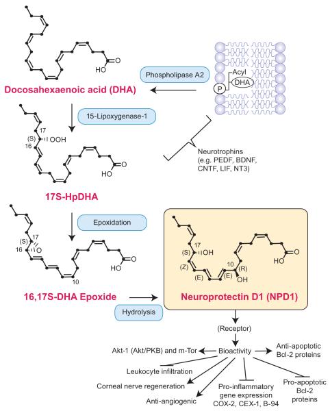
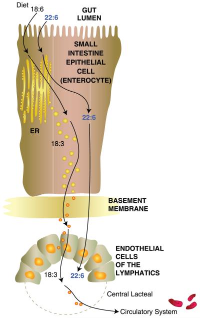
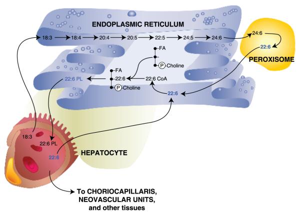
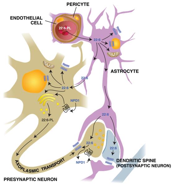
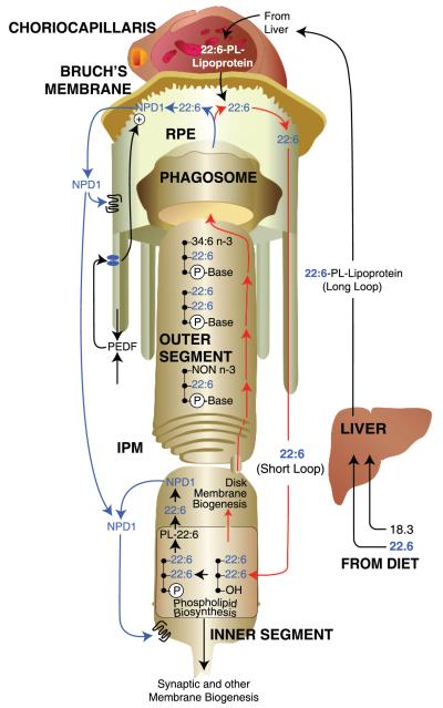
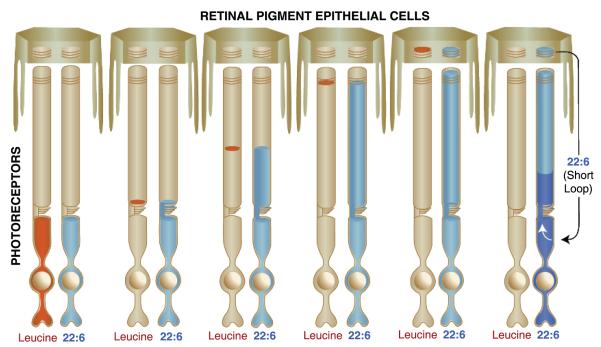
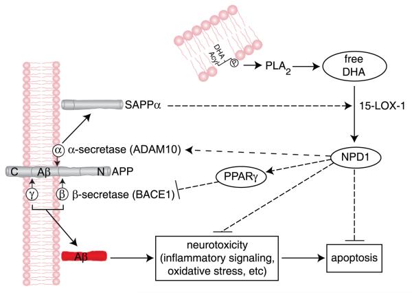
Essential polyunsaturated fatty acids (PUFAs) are critical nutritional lipids that must be obtained from the diet to sustain homeostasis. Omega-3 and -6 PUFAs are key components of biomembranes and play important roles in cell integrity, development, maintenance, and function. The essential omega-3 fatty acid family member docosahexaenoic acid (DHA) is avidly retained and uniquely concentrated in the nervous system, particularly in photoreceptors and synaptic membranes. DHA plays a key role in vision, neuroprotection, successful aging, memory, and other functions. In addition, DHA displays anti-inflammatory and inflammatory resolving properties in contrast to the proinflammatory actions of several members of the omega-6 PUFAs family. This review discusses DHA signalolipidomics, comprising the cellular/tissue organization of DHA uptake, its distribution among cellular compartments, the organization and function of membrane domains rich in DHA-containing phospholipids, and the cellular and molecular events revealed by the uncovering of signaling pathways regulated by DHA and docosanoids, the DHA-derived bioactive lipids, which include neuroprotectin D1 (NPD1), a novel DHA-derived stereoselective mediator. NPD1 synthesis agonists include neurotrophins and oxidative stress; NPD1 elicits potent anti-inflammatory actions and prohomeostatic bioactivity, is anti-angiogenic, promotes corneal nerve regeneration, and induces cell survival. In the context of DHA signalolipidomics, this review highlights aging and the evolving studies on the significance of DHA in Alzheimer's disease, macular degeneration, Parkinson's disease, and other brain disorders. DHA signalolipidomics in the nervous system offers emerging targets for pharmaceutical intervention and clinical translation.


Figures

Figure 1
Figure 1
Biosynthesis of neuroprotectin D1 (NPD1). A membrane phospholipid containing a docosahexaenoyl chain at sn-2 is hydrolyzed by phospholipase A2, generating free (unesterified) docosahexaenoic acid (DHA) (22:6). Lipoxygenation is then followed by epoxidation and hydrolysis, to generate NPD1. Thus far, a binding site for NPD1 has been identified in retinal pigment epithelium cells and polymorphonuclear cells (133). BDNF, brain-derived neurotrophic factor; CNTF, ciliary neurotrophic factor; CEX-1, cytokine exodus protein-1; COX-2, cyclooxygenase-2; LIF, leukemia inhibitory factor; NT3, neurotrophin 3; PEDF, pigment epithelium-derived factor.
Figure 2
Figure 2
Flow of the ω-3 fatty acid (FA), docosahexaenoic acid (DHA) (22:6), or its precursor, α-linolenic acid (ALA) (18:3), from the gastrointestinal lumen to the lymphatic system. DHA is required for the synthesis of phospholipids for nervous system membrane biogenesis. If unavailable, DHA can be synthesized from ALA. Enterocytes of the small intestine take up these FAs and package them for delivery to the endothelial cells of the lymphatic system. From there, they are transferred to the central lacteal and delivered to the circulatory system for transport. Detailed molecular events are not well understood as of yet.
Figure 3
Figure 3
Desaturation and elongation of α-linolenic acid (ALA) (18:3) within the hepatocyte. Systemic ALA is taken up by hepatocytes and transferred to the endoplasmic reticulum, where a series of desaturation and elongation steps occurs, leading to the formation of a 24-carbon, 6-double-bond FA [24:6, tetracosahexaenoic acid (THA)]. 24:6 is then conveyed to peroxisomes, converted to 22:6 by β-degradation, and delivered back to the endoplasmic reticulum. 22:6 (DHA) is then attached to the n-2 position of phosphatidyl choline to form a 22:6 phospholipid (22:6-PL), followed by release to the circulatory system for delivery to the choriocapillaris behind the retina and neovascular unit within the brain, as well as to other tissues.
Figure 4
Figure 4
Movement of docosahexaenoic acid (DHA) (22:6) through the neurovascular unit and its disposition within neurons and astrocytes. 22:6-phospholipids (22:6-PLs) are taken up by endothelial cells from the circulation and transferred to astrocytes within the central nervous system; from there, 22:6 is incorporated into astrocytes or transferred to neurons. 22:6 is also available for conversion to neuroprotectin D1 (NPD1) upon inductive signals. After packaging in the endoplasmic reticulum and the Golgi apparatus, 22:6 is also transported to the synaptic terminal to become incorporated into synaptic elements. Arrows represent possible routes. Molecular characterization of transporter(s) and receptors remains to be done.
Figure 5
Figure 5
Docosahexaenoic acid (DHA) (22:6) delivery to photoreceptors, photoreceptor membrane renewal, and synthesis of neuroprotectin D1 (NPD1). DHA (22:6) or immediate precursors are obtained by diet. Once within the liver, hepatocytes incorporate 22:6 into 22:6-phospholipid (22:6-PL)-lipoproteins, which are then transported to the choriocapillaris. 22:6 crosses Bruch’s membrane from the subretinal circulation and is taken up by the retinal pigment epithelium (RPE) cells lining the back of the retina to subsequently be sent to the underlying photoreceptors. This targeted delivery route from the liver to the retina is referred to as the 22:6 long loop. Then 22:6 passes through the interphotoreceptor matrix (IPM) and into the photoreceptor inner segment, where it is incorporated into phospholipids for cell membrane, organelles, and disk membrane biogenesis. As new 22:6-rich disks are synthesized at the base of the photoreceptor outer segment, older disks are pushed apically toward the RPE cells. Photoreceptor tips are phagocytized by the RPE cells each day, removing the oldest disks. The resulting phagosomes are degraded within the RPE cells, and 22:6 is recycled back to photoreceptor inner segments for new disk membrane biogenesis. This local recycling is referred to as the 22:6 short loop. Upon inductive signaling, such as pigment-epithelium derived factor (PEDF), 22:6 can be obtained from a phospholipid pool for the synthesis of neuroprotectin D1 (NPD1).
Figure 6
Figure 6
The incorporation of amino acids (e.g., leucine) and docosahexaenoic acid (DHA) during photoreceptor outer segment disk membrane biogenesis. Amino acids are utilized by photoreceptors in the synthesis of new outer segment disk membrane opsin and can be followed, when labeled with a tracking tag such as tritium, by autoradiography as they move into and through photoreceptor outer segments. The amino acid leucine (red), a component of the opsin protein, is avidly incorporated into disk membrane. However, because this amino acid is in great demand for many proteins, its photoreceptor pool is rapidly turned over. Initially, labeled leucine appears at the bottom of the outer segment, where the opsin protein is inserted into new membranes. A narrow band of label occurs as more membranes are synthesized, but labeling ceases as all labeled leucine is used up. The labeled band is slowly pushed apically as more membranes are synthesized, finally arriving at the photoreceptor tip in about 10 days in a warm-blooded animal. As the photoreceptor tip is phagocytized by the RPE cells, the labeled band appears within the RPE cell cytoplasm until degradation is complete. Leucine forms a tight band with no dispersion because the opsin molecule is locked into the structure of the disk membrane. Lipids do not follow this pattern. Labeled fatty acids (FAs) immediately disperse through the membranes of the outer segment, indicating that they exhibit free lateral and vertical movement (40). DHA (blue) is an exception. Once it enters the outer segment, it becomes integrated into the structure of the disk membrane and remains there until the disks are shed into the RPE cells. In addition, the pool of this FA is continually replenished (indicated by the white arrow) by recycling the 22:6 back to the inner segment from the retinal pigment epithelium, resulting in continual labeling of the new disk membranes. Importantly, the labeled front of the moving leucine band exactly matches the front of the 22:6 label. This occurs because the 22:6 becomes associated with the opsin molecule within the disk membrane and is thus, prevented from lateral and vertical free movement within the outer segment.
Figure 7
Figure 7
Mechanism for neuroprotectin D1 (NPD1) induction of nonamyloidogenic and neurotrophic bioactivity. Docosahexaenoic acid (DHA) (22:6) is excised by phospholipase A2 (PLA2) to yield free DHA; in turn, free DHA is 15-lipoxygenated to generate NPD1, which then activates neuroprotective signaling. These events are mediated, in part, by shifting beta-amyloid precursor protein (βAPP) processing from an amyloidogenic into a neurotrophic, nonamyloidogenic pathway and by inhibiting apoptosis, blocking inflammatory signaling, and promoting cell survival. Beta-site amyloid precursor protein-cleaving enzyme 1 (BACE1) (β-secretase-1) activity is suppressed, and α-secretase distintegrin and metalloproteinase 10 (ADAM10) activity is stimulated, thus down-regulating amyloid-beta 42 (Aβ42) peptide release. NPD1 signaling to BACE1 and ADAM10 may be mediated via other neuromolecular factors. The ADAM10 cleavage product soluble amyloid precursor protein alpha (sAPPα) further induces the conversion of free DHA into NPD1, thus constituting a positive, neurotrophic feedback loop.

References

    1. Agostoni C. Role of long-chain polyunsaturated fatty acids in the first year of life. J. Pediatr. Gastroenterol. Nutr. 2008;47:S41–44. - PubMed
    1. Akbar M, Calderon F, Wen Z, Kim H. Docosahexaenoic acid: a positive modulator of Akt signaling in neuronal survival. Proc. Natl. Acad. Sci. USA. 2005;102:10858–63. - PMC - PubMed
    1. Amor S, Puentes F, Baker D, van der Valk P. Inflammation in neurodegenerative diseases. Immunology. 2010;129:154–69. - PMC - PubMed
    1. Anders A, Gilbert S, Garten W, Postina R, Fahrenholz F. Regulation of the alpha-secretase ADAM10 by its prodomain and proprotein convertases. FASEB J. 2001;15:1837–39. - PubMed
    1. Anderson RE, Maude MB, McClellan M, Matthes MT, Yasumura D, LaVail MM. Low docosahexaenoic acid levels in rod outer segments of rats with P23H and S334ter rhodopsin mutations. Mol. Vis. 2002;8:351–58. - PubMed

Publication types

MeSH terms

Substances

LinkOut - more resources