. 2012 Jan;18(1-2):1-16.
doi: 10.1089/ten.TEA.2011.0301. Epub 2011 Sep 23.

Initial binding and recellularization of decellularized mouse lung scaffolds with bone marrow-derived mesenchymal stromal cells

Affiliations

Initial binding and recellularization of decellularized mouse lung scaffolds with bone marrow-derived mesenchymal stromal cells

Amanda B Daly et al. Tissue Eng Part A. 2012 Jan.

Abstract

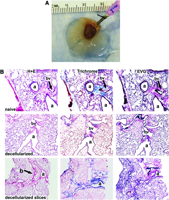
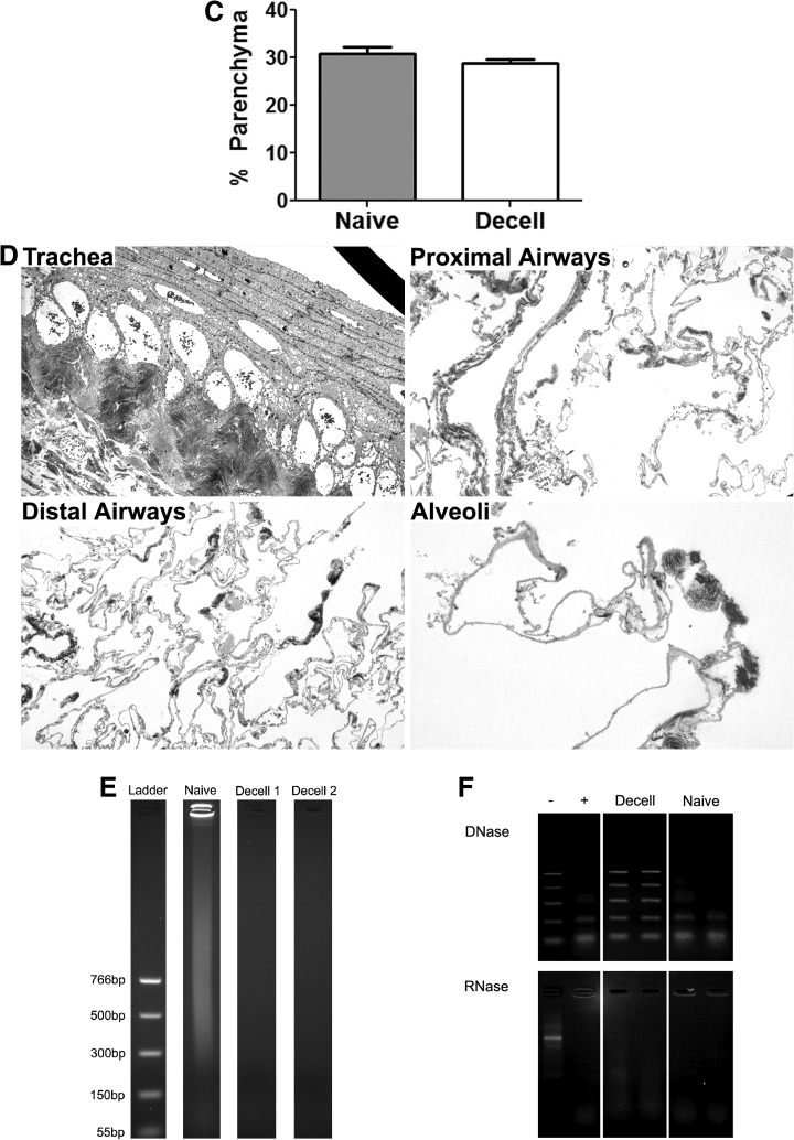
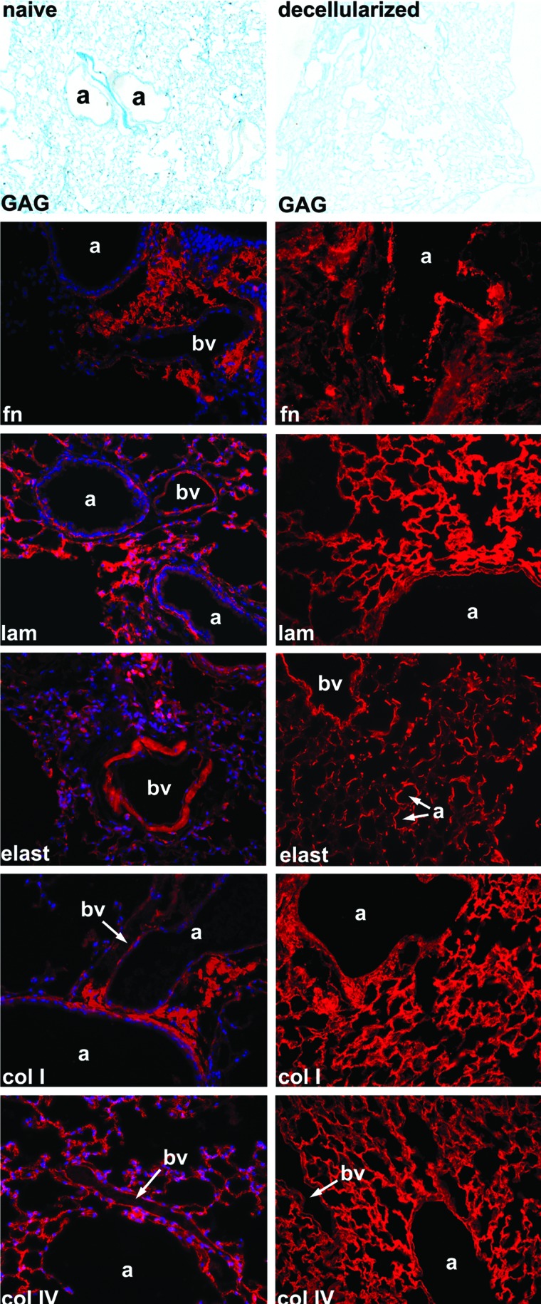
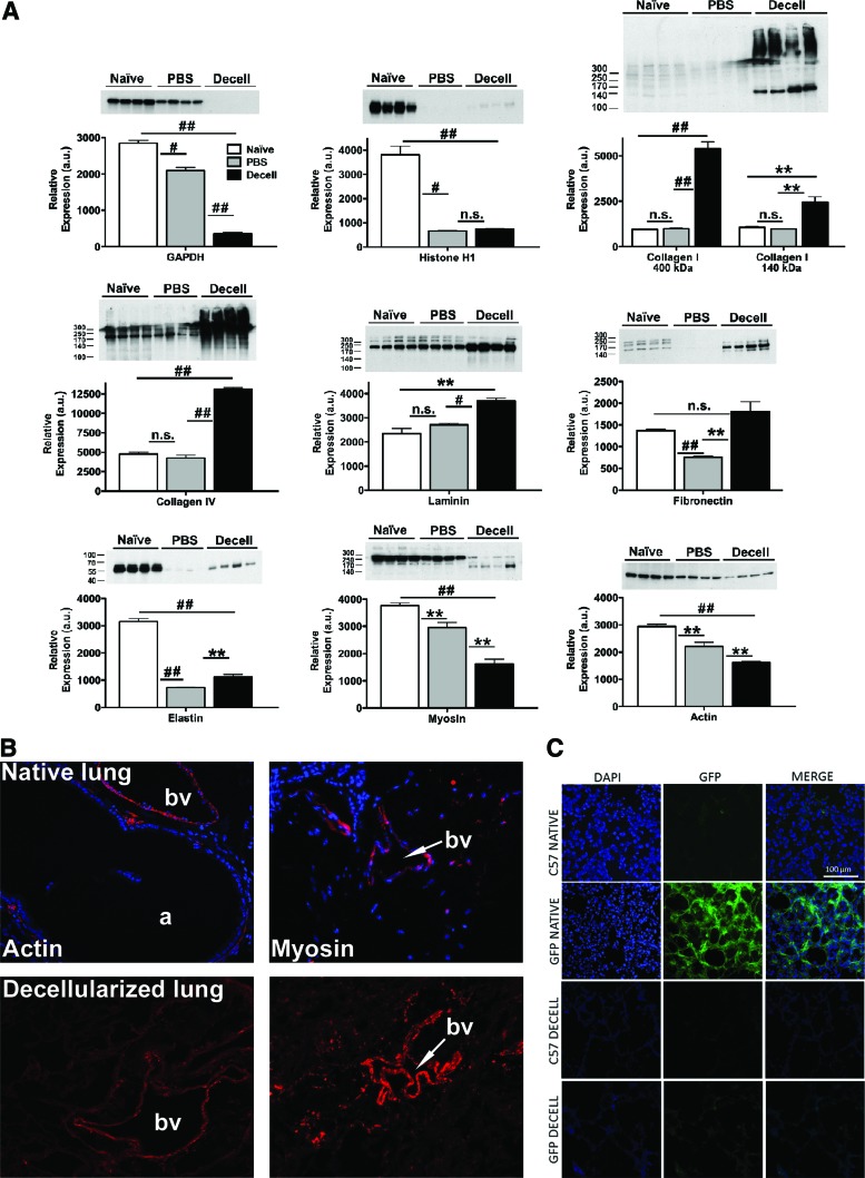
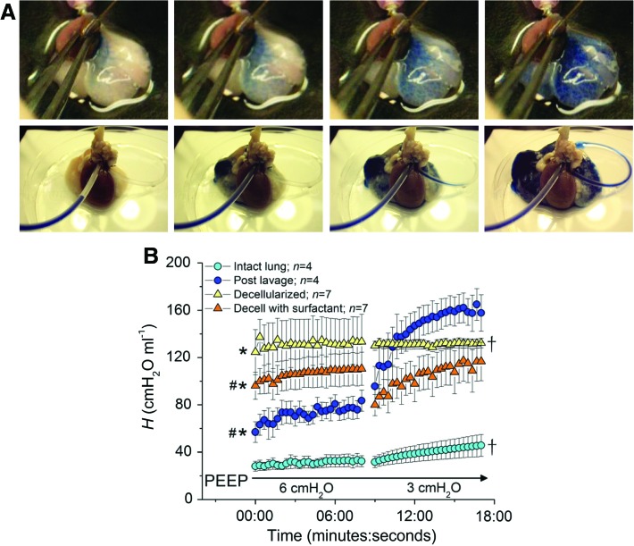
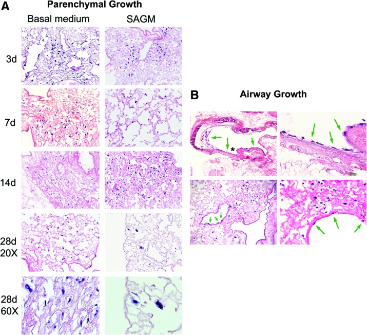
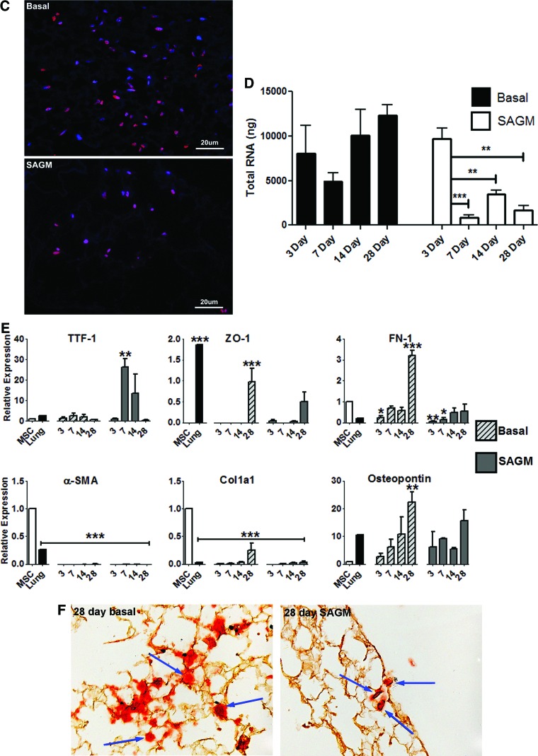
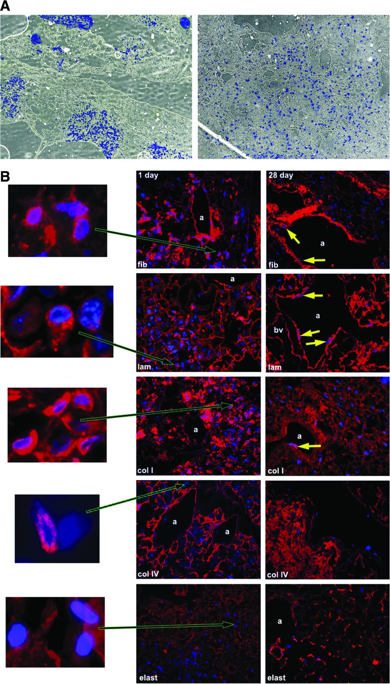
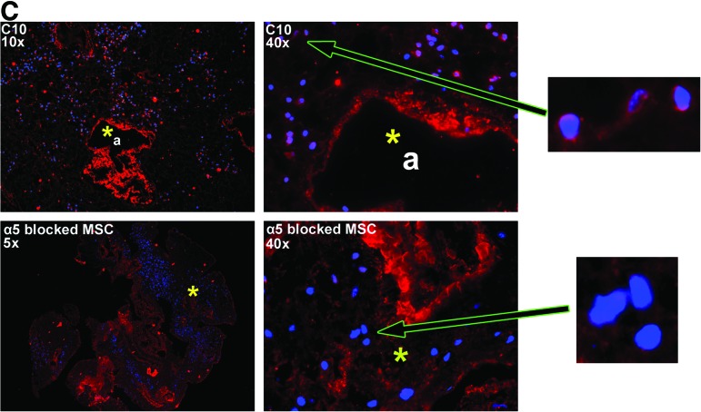
Recellularization of whole decellularized lung scaffolds provides a novel approach for generating functional lung tissue ex vivo for subsequent clinical transplantation. To explore the potential utility of stem and progenitor cells in this model, we investigated recellularization of decellularized whole mouse lungs after intratracheal inoculation of bone marrow-derived mesenchymal stromal cells (MSCs). The decellularized lungs maintained structural features of native lungs, including intact vasculature, ability to undergo ventilation, and an extracellular matrix (ECM) scaffold consisting primarily of collagens I and IV, laminin, and fibronectin. However, even in the absence of intact cells or nuclei, a number of cell-associated (non-ECM) proteins were detected using mass spectroscopy, western blots, and immunohistochemistry. MSCs initially homed and engrafted to regions enriched in types I and IV collagen, laminin, and fibronectin, and subsequently proliferated and migrated toward regions enriched in types I and IV collagen and laminin but not provisional matrix (fibronectin). MSCs cultured for up to 1 month in either basal MSC medium or in a small airways growth media (SAGM) localized in both parenchymal and airway regions and demonstrated several different morphologies. However, while MSCs cultured in basal medium increased in number, MSCs cultured in SAGM decreased in number over 1 month. Under both media conditions, the MSCs predominantly expressed genes consistent with mesenchymal and osteoblast phenotype. Despite a transient expression of the lung precursor TTF-1, no other airway or alveolar genes or vascular genes were expressed. These studies highlight the power of whole decellularized lung scaffolds to study functional recellularization with MSCs and other cells.


Figures

FIG. 1.
FIG. 1.
Histologic assessment of decellularized whole mouse lungs and lung slices demonstrates preservation of normal architecture. (A) Whole decellularized heart-lung bloc. The trachea is cannulated with a 18 gauge blunted needle. (B) H&E, Masson's Trichrome collagen, and Verhoeff's Van Gieson staining of native lungs, decellularized whole lungs, and approximately 1-mm-thick slices of decellularized lungs. Original magnifications: 100×. a, airway; bv, blood vessel. (C) Percent parenchyma in native versus decellularized lungs. Means ± standard error of three lungs assessed for each experimental condition are shown. (D) Transmission electron micrograph images of different regions of a representative decellularized whole mouse lung are shown. Original magnifications (A) 600×, (B) 1000×, (C) 1000×, (D) 3000×. (E) Total DNA content of a native C57Bl/6 mouse lung versus two representative decellularized whole lungs. (F) Demonstration of residual RNase but not DNAse activities in decellularized whole lungs. DNA and RNA ladders are shown in the first column (−). Ladders with addition of either DNAse or RNAse are shown in the second column as positive controls. Two different representative native lungs and decellularized lungs are shown for comparison. H&E, hematoxylin and eosin. Color images available online at www.liebertonline.com/tea
FIG. 1.
FIG. 1.
Histologic assessment of decellularized whole mouse lungs and lung slices demonstrates preservation of normal architecture. (A) Whole decellularized heart-lung bloc. The trachea is cannulated with a 18 gauge blunted needle. (B) H&E, Masson's Trichrome collagen, and Verhoeff's Van Gieson staining of native lungs, decellularized whole lungs, and approximately 1-mm-thick slices of decellularized lungs. Original magnifications: 100×. a, airway; bv, blood vessel. (C) Percent parenchyma in native versus decellularized lungs. Means ± standard error of three lungs assessed for each experimental condition are shown. (D) Transmission electron micrograph images of different regions of a representative decellularized whole mouse lung are shown. Original magnifications (A) 600×, (B) 1000×, (C) 1000×, (D) 3000×. (E) Total DNA content of a native C57Bl/6 mouse lung versus two representative decellularized whole lungs. (F) Demonstration of residual RNase but not DNAse activities in decellularized whole lungs. DNA and RNA ladders are shown in the first column (−). Ladders with addition of either DNAse or RNAse are shown in the second column as positive controls. Two different representative native lungs and decellularized lungs are shown for comparison. H&E, hematoxylin and eosin. Color images available online at www.liebertonline.com/tea
FIG. 2.
FIG. 2.
Immunofluorescent and histologic staining demonstrates retained ECM proteins in whole decellularized lungs. For each set of IF images, the specific ECM protein is shown in red with a DAPI nuclear costain. Glycosaminoglycan content is stained blue with Alcian blue stain. Fn, fibronectin; lam, laminin; elast, elastin; coll, type I collagen; colIV, type IV collagen; a, airway; bv, blood vessel. Representative photomicrographs from a decellularized C57Bl/6 mouse lung are shown. Original magnification 400× for IF photomicrographs and 100× for Alcian blue photomicrographs. ECM, extracellular matrix; IF, immunofluorescent; DAPI, 4′,6′-diamidino-2-phenylindane. Color images available online at www.liebertonline.com/tea
FIG. 3.
FIG. 3.
Intracellular proteins can be detected in decellularized lung homogenates. (A) Four representative blots are shown, respectively, for native lungs, lungs sham decellularized with PBS only, and for lungs undergoing full decellularization. Quantitative data are presented as mean±SEM. Significant interactions were identified by one-way analysis of variance with Bonferroni post hoc analysis using Prism software. *p≤0.05, #p≤0.001, ##p≤0.0001, **p≤0.01, n.s., non-significant. (B) Representative immunofluorescent staining for smooth muscle actin and smooth muscle myosin heavy chain. Original magnifications=200×. (C) Direct GFP fluorescence in native and decellularized C57Bl/6 and eGFP-expressing mouse lungs. Original magnifications 200×. PBS, phosphate-buffered saline; SEM, standard error of the mean; eGFP, enhanced green fluorescent protein. Color images available online at www.liebertonline.com/tea
FIG. 4.
FIG. 4.
Vascular perfusion is preserved and lung elastance increased in whole decellularized mouse lungs. (A) Sequential images of dye perfusion in mouse (top panel) and rat (bottom panel) lungs. (B) Mean values for lung elastance (±SEM) measured at PEEP of 6 and 3 cm H2O, plotted against time, in intact native lungs, then after saline lavage, and then after decellularization and intratracheal instillation of surfactant (into decellularized lungs). * indicates that baseline mean lung elastance is significantly greater than that of the intact lung (p≤0.05), # indicates that baseline mean lung elastance is significant less than that of the decellularized lung (p≤0.05), † indicates that ΔH is significantly less than that of the lavaged lung (p≤0.05). PEEP, positive end-expiratory pressure. Color images available online at www.liebertonline.com/tea
FIG. 5.
FIG. 5.
Intratracheally inoculated MSCs cultured up to 1 month in both basal MSC media and in SAGM grow in parenchymal and airway regions of decellularized whole mouse lungs. Representative photomicrographs depict MSCs in parenchymal lung regions (A) and in airways (B). High power images demonstrate several morphologies of MSCs growing in parenchymal lung regions for 7 days in basal medium (top row) or 14 days in SAGM (bottom row) (A). Green arrows highlight cells growing in airways, and the asterisk in the upper left-hand image show the region magnified in the upper right-hand image. Original magnifications in (A) are 200×, except 600× where indicated by label “60×”; beginning in upper left-hand corner and proceeding clockwise. Original magnifications in (B) are 100×, 400×, 400×, and 200×. (C) Ki67 immunofluorescence (red) of MSCs cultured in either basal MSC medium or in SAGM demonstrates widespread expression in the majority of cells 14 days after intratracheal inoculation into decellularized mouse lungs. Representative photomicrographs are shown with DAPI nuclear costaining (original magnification 400×). (D) Total RNA in homogenates of whole decellularized lungs increases over time after inoculation with MSCs and culture in basal MSC media but decreases with culture in SAGM. Values represent means±standard error of the means of three lungs evaluated for each experimental condition. *p≤0.05, **p≤0.005, ***p≤0.0005. (E) Relative mRNA expression for selected genes is depicted. Values represent means±standard error of the means of three lungs per experimental condition. *p≤0.05, **p≤0.005, ***p≤0.0005. (F) Alizarin red staining of MSCs cultured in either basal MSC medium or in SAGM for 28 days after inoculation into whole decellularized mouse lings. Blue arrows highlight individual cells. MSCs, mesenchymal stromal cells; SAGM, small airways growth media. Color images available online at www.liebertonline.com/tea
FIG. 5.
FIG. 5.
Intratracheally inoculated MSCs cultured up to 1 month in both basal MSC media and in SAGM grow in parenchymal and airway regions of decellularized whole mouse lungs. Representative photomicrographs depict MSCs in parenchymal lung regions (A) and in airways (B). High power images demonstrate several morphologies of MSCs growing in parenchymal lung regions for 7 days in basal medium (top row) or 14 days in SAGM (bottom row) (A). Green arrows highlight cells growing in airways, and the asterisk in the upper left-hand image show the region magnified in the upper right-hand image. Original magnifications in (A) are 200×, except 600× where indicated by label “60×”; beginning in upper left-hand corner and proceeding clockwise. Original magnifications in (B) are 100×, 400×, 400×, and 200×. (C) Ki67 immunofluorescence (red) of MSCs cultured in either basal MSC medium or in SAGM demonstrates widespread expression in the majority of cells 14 days after intratracheal inoculation into decellularized mouse lungs. Representative photomicrographs are shown with DAPI nuclear costaining (original magnification 400×). (D) Total RNA in homogenates of whole decellularized lungs increases over time after inoculation with MSCs and culture in basal MSC media but decreases with culture in SAGM. Values represent means±standard error of the means of three lungs evaluated for each experimental condition. *p≤0.05, **p≤0.005, ***p≤0.0005. (E) Relative mRNA expression for selected genes is depicted. Values represent means±standard error of the means of three lungs per experimental condition. *p≤0.05, **p≤0.005, ***p≤0.0005. (F) Alizarin red staining of MSCs cultured in either basal MSC medium or in SAGM for 28 days after inoculation into whole decellularized mouse lings. Blue arrows highlight individual cells. MSCs, mesenchymal stromal cells; SAGM, small airways growth media. Color images available online at www.liebertonline.com/tea
FIG. 6.
FIG. 6.
MSCs inoculated into decellularized mouse lungs initially localize to regions enriched in fibronectin, laminin, type I collagen, and type IV collagen but do not proliferate in fibronectin-rich regions after 28 days in culture. (A) MSCs are localized in clumps 1 day after intratracheal inoculation but spread throughout the parenchyma and along airways 28 days after culture in basal MSC medium. Representative colorized phase contrast images are shown. Original magnification 50×. (B) Representative photomicrographs obtained 1 and 28 days after MSC inoculation into decellularized whole mouse lungs. For each panel, specific ECM protein immunofluorescence is depicted in red with DAPI nuclear costaining. Enlargements demonstrate that MSCs also produce fibronectin, laminin, and collagen I but not collagen IV or elastin after either 1 or 28 days in culture. (C) Representative photomicrographs demonstrating a different pattern of initial localization of inoculated C10 mouse lung epithelial cells with respect to fibronectin as compared to MSCs. Further, blocking the α5 integrin on MSCs with a neutralizing antibody prior to cell inoculation also results in MSCs localizing in areas not enriched in fibronectin. Enlargements demonstrate that the C10 cells themselves may produce small amounts of fibronectin whereas MSCs incubated with the neutralizing antibody do not appear to make fibronectin. a, airways; bv, blood vessels; fn, fibronectin; lam, laminin; elast, elastin; colI, type I collagen; colIV, type IV collagen. Yellow arrows depict MSCs growing along airway walls. Original magnifications 400×. Color images available online at www.liebertonline.com/tea
FIG. 6.
FIG. 6.
MSCs inoculated into decellularized mouse lungs initially localize to regions enriched in fibronectin, laminin, type I collagen, and type IV collagen but do not proliferate in fibronectin-rich regions after 28 days in culture. (A) MSCs are localized in clumps 1 day after intratracheal inoculation but spread throughout the parenchyma and along airways 28 days after culture in basal MSC medium. Representative colorized phase contrast images are shown. Original magnification 50×. (B) Representative photomicrographs obtained 1 and 28 days after MSC inoculation into decellularized whole mouse lungs. For each panel, specific ECM protein immunofluorescence is depicted in red with DAPI nuclear costaining. Enlargements demonstrate that MSCs also produce fibronectin, laminin, and collagen I but not collagen IV or elastin after either 1 or 28 days in culture. (C) Representative photomicrographs demonstrating a different pattern of initial localization of inoculated C10 mouse lung epithelial cells with respect to fibronectin as compared to MSCs. Further, blocking the α5 integrin on MSCs with a neutralizing antibody prior to cell inoculation also results in MSCs localizing in areas not enriched in fibronectin. Enlargements demonstrate that the C10 cells themselves may produce small amounts of fibronectin whereas MSCs incubated with the neutralizing antibody do not appear to make fibronectin. a, airways; bv, blood vessels; fn, fibronectin; lam, laminin; elast, elastin; colI, type I collagen; colIV, type IV collagen. Yellow arrows depict MSCs growing along airway walls. Original magnifications 400×. Color images available online at www.liebertonline.com/tea

References

    1. Bhatia S.K. Tissue engineering for clinical applications. Biotechnol J. 2010;5:1309. - PubMed
    1. Ott H.C. Matthiesen T.S. Goh S.K. Black L.D. Kren S.M. Netoff T.I. Taylor D.A. Perfusion-decellularized matrix: using nature's platform to engineer a bioartificial heart. Nature Med. 2008;14:213. - PubMed
    1. Uygun B.E. Soto-Gutierrez A. Yagi H. Izamis M.L. Guzzardi M.A. Shulman C. Milwid J. Kobayashi N. Tilles A. Berthiaume F. Hertl M. Nahmias Y. Yarmush M.L. Uygun K. Organ reengineering through development of a transplantable recellularized liver graft using decellularized liver matrix. Nat Med. 2010;16:814. - PMC - PubMed
    1. Mondrinos M.J. Koutzaki S. Lelkes P.I. Finck C.M. A tissue-engineered model of fetal distal lung tissue. Am J Physiol Lung Cell Mol Physiol. 2007;293:L639. - PubMed
    1. Lin Y.M. Boccaccini A.R. Polak J.M. Bishop A.E. Maquet V. Biocompatibility of poly-DL-lactic acid (PDLLA) for lung tissue engineering. J Biomater Appl. 2005;21:109. - PubMed

MeSH terms

LinkOut - more resources