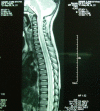
Spinal epidural abscess with myelitis and meningitis caused by Streptococcus pneumoniae in a young child
- PMID: 21756576
- PMCID: PMC3127372
- DOI: 10.1179/107902610x12883422813507
Spinal epidural abscess with myelitis and meningitis caused by Streptococcus pneumoniae in a young child
Abstract
Background: Spinal epidural abscess (SEA) in children is a rare infectious emergency warranting prompt intervention. Predisposing factors include immunosuppression, spinal procedures, and local site infections such as vertebral osteomyelitis and paraspinal abscess. Staphylococcus aureus is the most common isolate.
Design: Case report and literature review.
Findings: A 2.5-year-old boy with tetraparesis was found to have an SEA in the posterior lumbar epidural space with evidence of meningitis and myelitis on MRI spine in the absence of any local or systemic predisposing factors or spinal procedures. Streptococcus pneumoniae was isolated from the evacuated pus.
Conclusions: Definitive treatment of SEA is a combination of surgical decompression and iv antibiotics. Timely management limits the extent of neurological deficit.
Figures


References
-
- Enberg RN, Kaplan RJ. Spinal epidural abscess in children: early diagnosis and immediate surgical drainage is essential to forestall paralysis. Clin Pediatr 1974;13(3):247–53 - PubMed
-
- Auletta JJ, John CC. Spinal epidural abscesses in children: a 15-year experience and review of the literature. Clin Infect Dis 2001;32(1):9–16 - PubMed
-
- Darouiche RO. Spinal epidural abscess. N Engl J Med 2006;355(19):2012–20 - PubMed
-
- Akalan N, Ozgen T. Infection as a cause of spinal cord compression: a review at 36 spinal epidural abscess cases. Acta Neurochir (Wien) 2000;142(1):17–21 - PubMed
-
- Phillips JMG, Stedeford JC, Hartsilver E, Roberts C. Epidural abscess complicating insertion of epidural catheters. Br J Anaesth 2002;89(5):778–82 - PubMed
Publication types
MeSH terms
LinkOut - more resources
Full Text Sources
Medical
