. 2012 Jan;56(1):137-45.
doi: 10.1016/j.jhep.2011.05.025. Epub 2011 Jul 12.

Sphingosine kinase-2 inhibition improves mitochondrial function and survival after hepatic ischemia-reperfusion

Affiliations

Sphingosine kinase-2 inhibition improves mitochondrial function and survival after hepatic ischemia-reperfusion

Yanjun Shi et al. J Hepatol. 2012 Jan.

Abstract

Background & aims: The mitochondrial permeability transition (MPT) and inflammation play important roles in liver injury caused by ischemia-reperfusion (IR). This study investigated the roles of sphingosine kinase-2 (SK2) in mitochondrial dysfunction and inflammation after hepatic IR.

Methods: Mice were gavaged with vehicle or ABC294640 (50 mg/kg), a selective inhibitor of SK2, 1 h before surgery and subjected to 1 h-warm ischemia to ~70% of the liver followed by reperfusion.

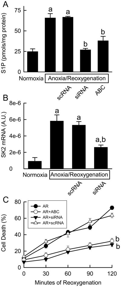
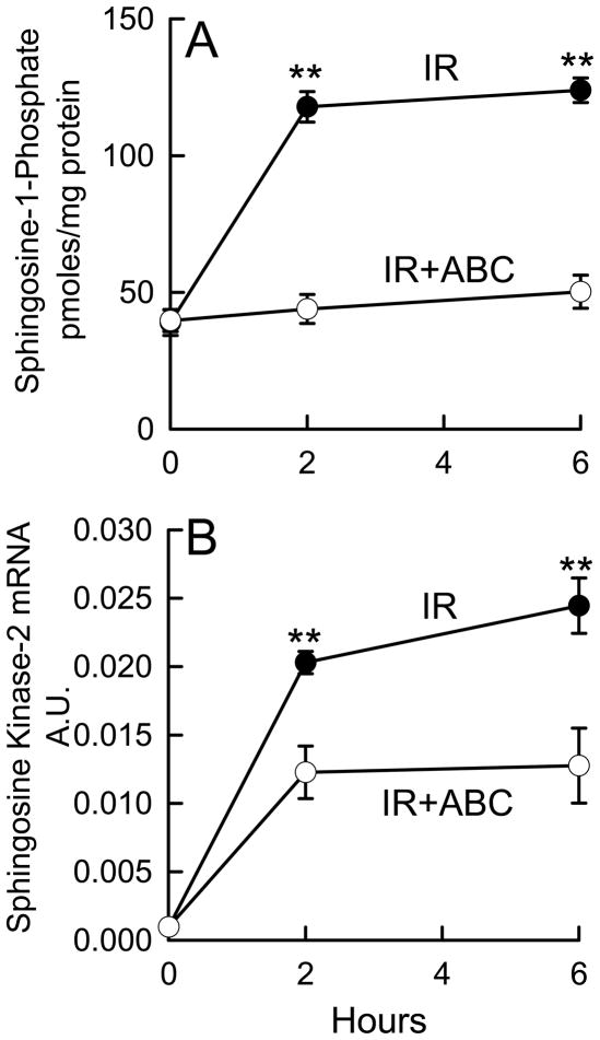
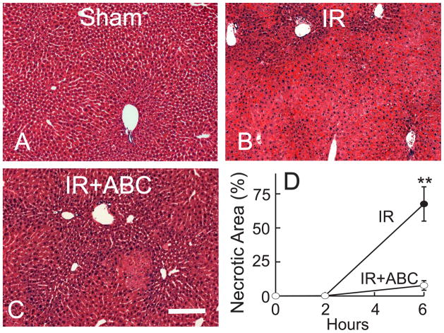
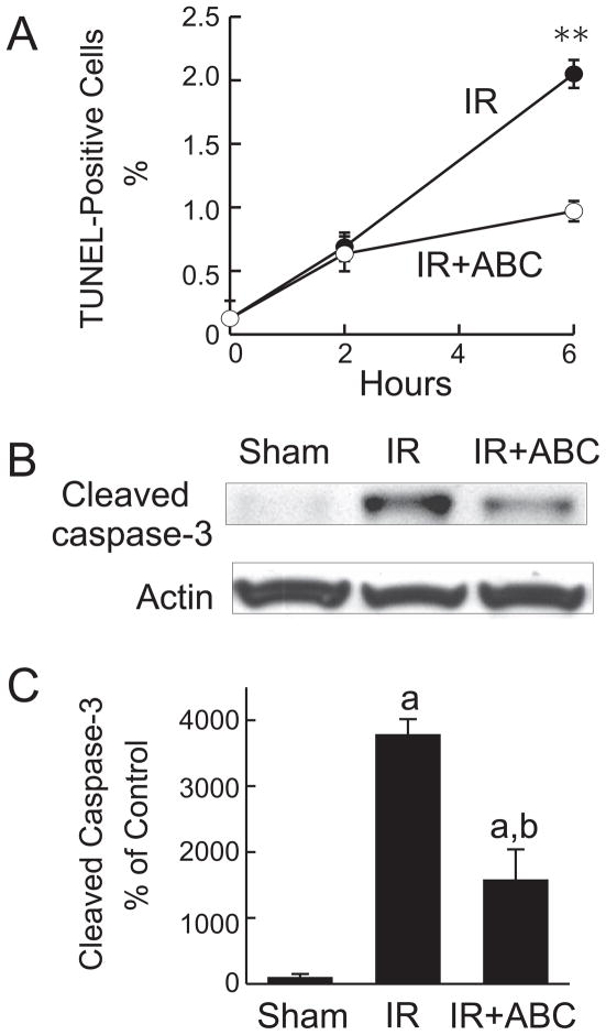
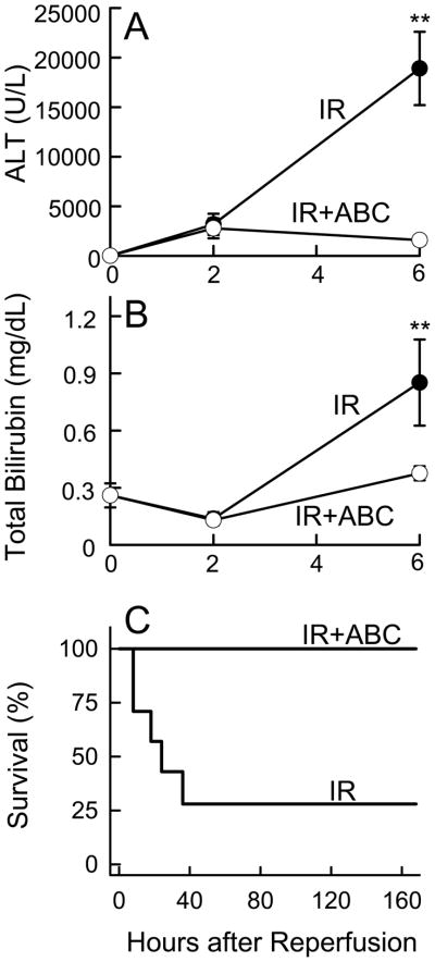
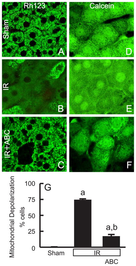
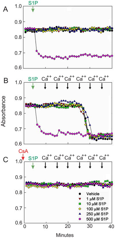
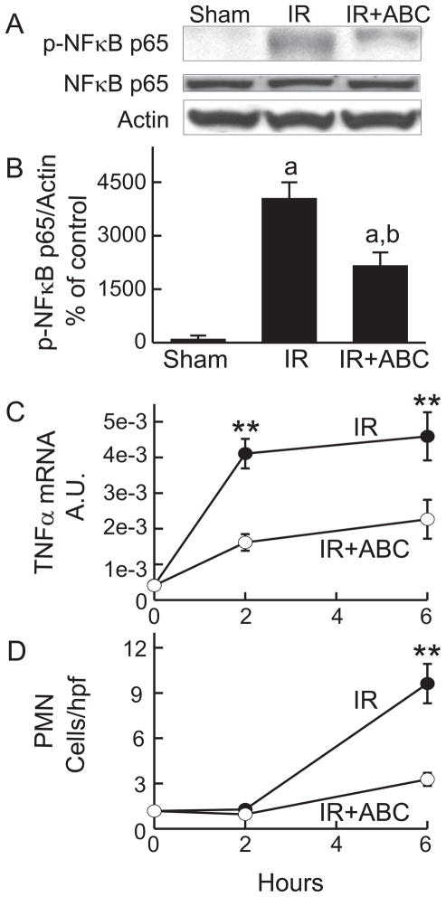
Results: Following IR, hepatic SK2 mRNA and sphingosine-1-phosphate (S1P) levels increased ~25- and 3-fold, respectively. SK2 inhibition blunted S1P production and liver injury by 54-91%, and increased mouse survival from 28% to 100%. At 2 h after reperfusion, mitochondrial depolarization was observed in 74% of viable hepatocytes, and mitochondrial voids excluding calcein disappeared, indicating MPT onset in vivo. SK2 inhibition decreased mitochondrial depolarization and prevented MPT onset. Inducible nitric oxide synthase, phosphorylated NFκB-p65, TNFα mRNA, and neutrophil infiltration, all increased markedly after hepatic IR, and these increases were blunted by SK2 inhibition. In cultured hepatocytes, anoxia/re-oxygenation resulted in increases of SK2 mRNA, S1P levels, and cell death. SK2 siRNA and ABC294640 each substantially decreased S1P production and cell death in cultured hepatocytes.

Conclusions: SK2 plays an important role in mitochondrial dysfunction, inflammation responses, hepatocyte death, and survival after hepatic IR and represents a new target for the treatment of IR injury.


Figures

Fig. 1
Fig. 1. ABC294640 and SK2 siRNA prevent anoxia/reoxygenation-induced cell death of cultured hepatocytes
Cultured hepatocytes were treated with ABC294640 and SK2 siRNA as indicated in “METHODS.” At 120 min after reoxygenation, S1P in hepatocytes was quantified by ELISA (A). SK2 mRNA was measured by real-time PCR (B). Cell death was detected by propidium iodide fluorescence at the indicated times (C). A.U., arbitrary unit; scRNA, scrambled RNA; siRNA, small interfering RNA. a, p<0.05 vs normoxia; b, p<0.05 vs anoxia/reoxygenation (n = 4 per group).
Fig. 2
Fig. 2. ABC294640 prevents upregulation of SK2 and S1P production after hepatic IR
Livers were harvested at 2 and 6 h after reperfusion. Sphingosine-1-phosphate (S1P) in liver tissues was quantified by ELISA (A). SK2 mRNA was measured by real time-PCR (B). IR, ischemia/reperfusion; ABC, ABC294640; A.U., arbitrary units. **, p<0.01 vs the ABC-treated group at the corresponding time point (n = 4 per group).
Fig. 3
Fig. 3. ABC294640 attenuates necrosis after hepatic IR
Livers were harvested at 2 and 6 h after reperfusion (IR), and liver slices were stained with H+E. Representative images at 6 h are shown (A to C). The bar is 100 μm. Necrotic areas were quantified by image analysis of 10 randomly selected fields (D). **, p<0.01 vs the ABC-treated group at the corresponding time point (n = 4 per group).
Fig. 4
Fig. 4. ABC294640 decreases apoptosis after hepatic IR
Livers were harvested at 2 and 6 h after reperfusion (IR). TUNEL-positive cells were counted in 10 randomly selected fields (A). **, p<0.01 vs the ABC-treated group at the corresponding time point. Representative Western blot images of cleaved caspase-3 are shown in B, and quantified by densitometry (C). a, p<0.05 vs sham-operation; b, p<0.05 vs IR (n = 4 per group).
Fig. 5
Fig. 5. ABC294640 protects liver function and improves survival after hepatic IR
Blood samples were collected at 2 and 6 h after reperfusion for ALT (A) and bilirubin measurement (B). **, p<0.01 vs the ABC-treated group at the corresponding time point (n = 4 per group). Survival probabilities (C) after IR were significantly different between vehicle- (n = 7) and ABC294640-treated mice (n = 9) by the Kaplan-Meier test.
Fig. 6
Fig. 6. ABC294640 prevents mitochondrial depolarization and onset of the MPT after hepatic IR
At 2 h after sham-operation or reperfusion, Rh123 plus PI or calcein-AM alone was infused, and intravital multiphoton microscopy was performed. Representative images of Rh123 are shown in A to C, and images of calcein in D to F. Rows are: upper, sham operation (sham); middle, ischemia-reperfusion (IR); lower, IR plus ABC294640-treatment. Bars are 5 μm. Viable cells with depolarized mitochondria were counted in 10 random fields for each liver (G). a, p<0.05 vs sham-operation; b, p<0.05 vs IR (n = 5 in the IR group and n = 4 in the sham and IR+ABC group, respectively). The majority of the cells in E show the MPT except the cell in the left upper corner (identified by the arrow), where incomplete MPT with some dark voids remaining is seen.
Fig. 7
Fig. 7. Effects of Sphingosine-1-Phosphate on the Mitochondrial Permeability Transition in Isolated Rat Liver Mitochondria
Mitochondrial swelling was assessed as a decrease in absorbance at 540 nm. A: Mitochondrial suspensions were treated with 0 to 500 μM S1P in the absence of cyclosporine A (CsA). B: After mitochondrial suspensions were treated with 0 to 500 μM S1P, aliquots of 50 μM CaCl2 were added every 5 minutes to induce the MPT. C: Mitochondrial suspensions were treated with 0 to 500 μM S1P in the presence of 2 μM CsA. Green arrows, addition of the vehicle or S1P; black arrows: addition of CaCl2; red arrow, addition of CsA. For all experiments, the response to 500 μM S1P is indicated by the pink circles. Shown are representative OD values of 4 independent experiments.
Fig. 8
Fig. 8. ABC294640 blunts upregulation of iNOS after hepatic IR
Livers were harvested at 2 and 6 h after ischemia/reperfusion (IR). Levels of iNOS and actin were determined by Western blotting. Blot images were shown in A. Images of iNOS were quantified by densitometry (B). **, p<0.01 vs the ABC-treated group at the corresponding time point (n = 4 per group).
Fig. 9
Fig. 9. ABC294640 blunts NF-κ B activation, TNFα mRNA increase and polymorphonuclear neutrophil infiltration after hepatic IR
Livers were harvested at 2 and 6 h after ischemia/reperfusion (IR). Western blot images of NF-κB p65 and phosphorylated NF-κB p65 at 2 h are shown in A and quantified by densitometry (B). TNFα mRNA in liver tissue was measured by quantitative real-time PCR (C). A.U., arbitrary units. MPO-positive cells detected by immunohistochemistry were counted in a blinded manner in 10 randomly selected fields using a 40x objective lens (D). **, p<0.01 vs the ABC-treated group at the corresponding time point (n = 4 per group).

Similar articles

Cited by

References

    1. Morales A, Lee H, Goni FM, Kolesnick R, Fernandez-Checa JC. Sphingolipids and cell death. Apoptosis. 2007 May;12(5):923–939. - PubMed
    1. Hait NC, Oskeritzian CA, Paugh SW, Milstien S, Spiegel S. Sphingosine kinases, sphingosine 1-phosphate, apoptosis and diseases. Biochim Biophys Acta. 2006 Dec;1758(12):2016–2026. - PubMed
    1. Xia P, Gamble JR, Rye KA, Wang L, Hii CS, Cockerill P, et al. Tumor necrosis factor-alpha induces adhesion molecule expression through the sphingosine kinase pathway. Proc Natl Acad Sci U S A. 1998 Nov 24;95(24):14196–14201. - PMC - PubMed
    1. Olivera A, Spiegel S. Sphingosine-1-phosphate as second messenger in cell proliferation induced by PDGF and FCS mitogens. Nature. 1993 Oct 7;365(6446):557–560. - PubMed
    1. Lai WQ, Irwan AW, Goh HH, Howe HS, Yu DT, Valle-Onate R, et al. Anti-inflammatory effects of sphingosine kinase modulation in inflammatory arthritis. J Immunol. 2008 Dec 1;181(11):8010–8017. - PubMed

Publication types

MeSH terms

Substances