. 2012 Feb;56(2):359-66.
doi: 10.1016/j.jhep.2011.05.023. Epub 2011 Jul 12.

HO-1-STAT3 axis in mouse liver ischemia/reperfusion injury: regulation of TLR4 innate responses through PI3K/PTEN signaling

Affiliations

HO-1-STAT3 axis in mouse liver ischemia/reperfusion injury: regulation of TLR4 innate responses through PI3K/PTEN signaling

Bibo Ke et al. J Hepatol. 2012 Feb.

Abstract

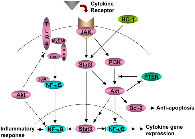
Background & aims: Signal transducer and activator of transcription 3 (STAT3), a key mediator of anti-inflammatory cytokine signaling, is essential for heme oxygenase-1 (HO-1)-induced cytoprotection. The phosphoinositide 3-kinase (PI3K)/phosphatase and tensin homolog delete on chromosome 10 (PTEN) pathways regulate diverse innate immune responses. This study was designed to investigate the role of STAT3 in the regulation of PI3K/PTEN cascade after HO-1 induction in a mouse model of innate immune-dominated liver ischemia/reperfusion injury (IRI).

Methods: Partial warm ischemia was produced in the left and middle hepatic lobes of C57BL/6 mice for 90 min, followed by 6h of reperfusion.

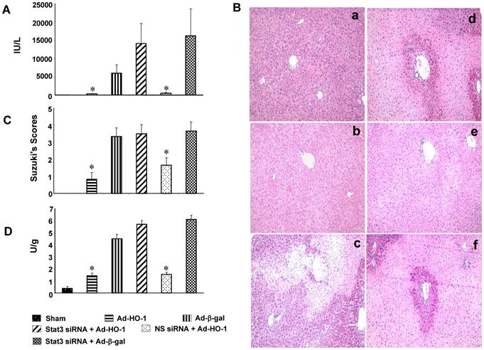
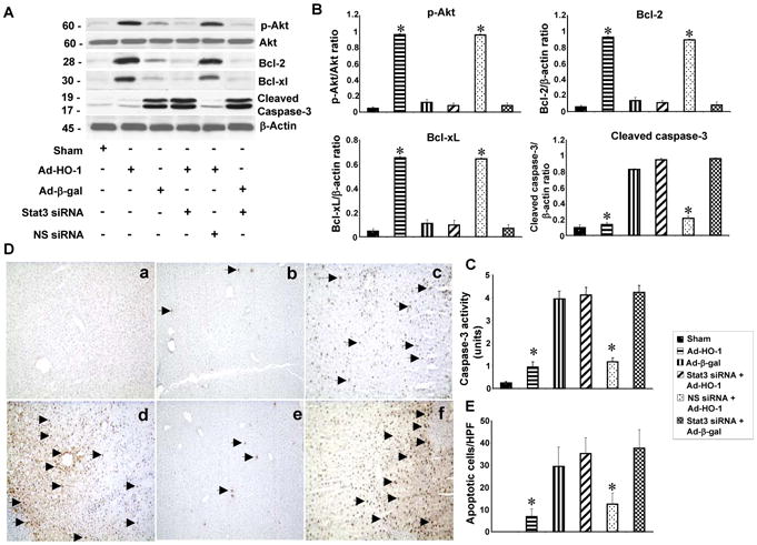
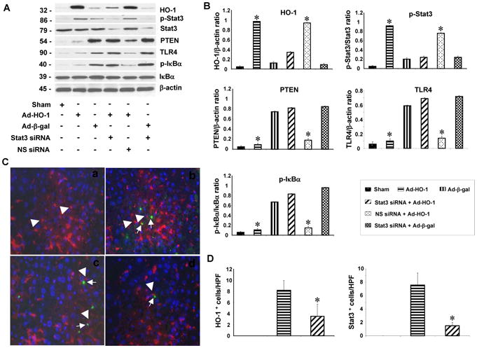
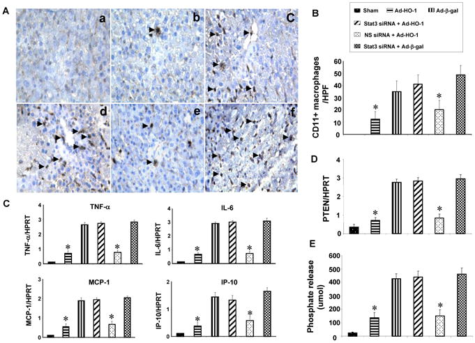
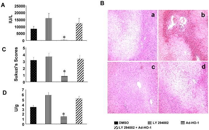
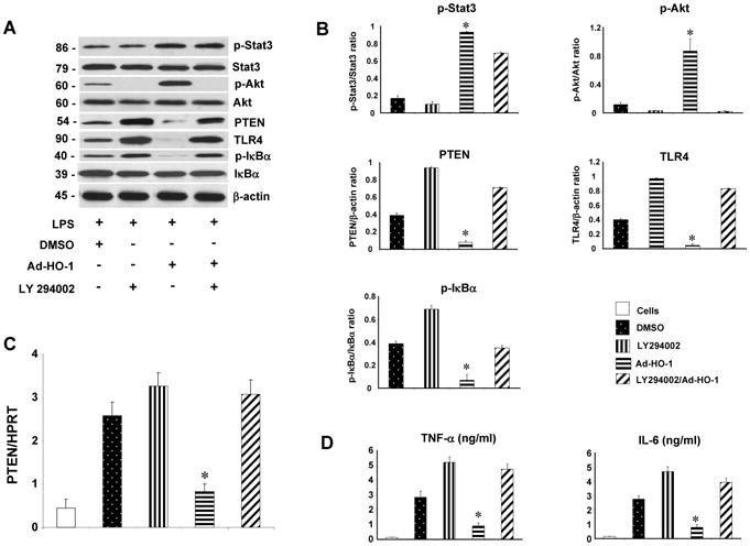
Results: Mice subjected to Ad-HO-1 transfer were resistant to liver IRI, and this cytoprotective effect correlated with increased intrahepatic PI3K/Akt and diminished PTEN expression. In contrast, mice undergoing adjunctive Ad-HO-1 treatment and STAT3 knockdown (siRNA) remained susceptible to IR-mediated local inflammatory response and hepatocellular damage. Consistent with decreased cell apoptosis and inhibited TLR4 expression after PI3K/Akt activation, treatment with specific PI3k inhibitor increased local inflammation and recreated liver IRI despite Ad-HO-1 gene transfer. Parallel in vitro studies with bone marrow derived-macrophages have confirmed that HO-1-STAT3 axis-induced PI3K/Akt negatively regulated PTEN expression in TLR4-dependent fashion.

Conclusions: These findings underscore the role of HO-1 induced STAT3 in modulating PI3K/PTEN in liver IRI cascade. Activating PI3K/Akt provides negative feedback mechanism for TLR4-driven inflammation. Identifying molecular pathways of STAT3 modulation in the innate immune system provides the rationale for novel therapeutic approaches for the management of liver inflammation and IRI in transplant patients.


Figures

Figure 1
Figure 1
HO-1-STAT3 signaling reduces severity of liver IRI. Mice were subjected to 90min of partial liver warm ischemia, followed by 6h reperfusion. (A). Hepatocellular function evaluated by sGPT (IU/L). Mean±SD (n=4-6 samples/group). *p<0.05. (B/C). The severity of liver IRI by Suzuki's histological grading: (a) Sham; (b) Ad-HO-1 (0.83±0.4); (c) Ad-β-gal (3.33±0.52); (d) siSTAT3+Ad-HO-1 (3.5±0.55); (e) nonspecific siRNA+Ad-HO-1 (1.2±0.41); (f) siSTAT3+Ad-β-gal (3.6±0.51). Representative of 4-6 mice/group; original magnification ×200. (D). Neutrophil accumulation analyzed by MPO activity (U/gm). Mean±SD (n=4-6 samples/group). *p< 0.05.
Figure 2
Figure 2
HO-1-STAT3 axis activates hepatic PI3K/Akt signaling and reduces IR-apoptosis. (A/B) Western-assisted analysis of phos-Akt, Bcl-2, Bcl-xl and cleaved caspase-3. Representative of three experiments. (C). Caspase-3 activity. Mean±SD; n=4-6 samples/group. *p<0.001. (D/E). Liver apoptosis by TUNEL staining. (a) Sham; (b) Ad-HO-1; (c) Ad-β-gal; (d) siSTAT3+Ad-HO-1; (e) nonspecific siRNA+Ad-HO-1; (f) siSTAT3+Ad-β-gal. Apoptotic cells are marked (arrow). Results scored semi-quantitatively by averaging the number of apoptotic cells (mean±SD) per field at 200×magnification (representative of 4-6 mice/group).
Figure 3
Figure 3
HO-1-STAT3 signaling reduces IR-induced TLR4 and NF-κB expression. (A/B) Western-assisted detection of HO-1, phos-STAT3, PTEN, TLR4, and phos-IκBα. Representative of three experiments. (C/D). Immunofluorescence staining of HO-1 and phos-STAT3 in ischemic livers. (a) Sham; (b) HO-1 expression in Ad-HO-1 treated livers; (c) Phos-STAT3 expression in Ad-HO-1 treated livers; (d) Phos-STAT3 expression in siSTAT3 treated livers. Note; positive staining in macrophages (head arrow), detected with CD68 mAb. Green: HO-1 and p-STAT3; Red: macrophage marker; Blue: DAPI nuclear stain. Results scored semi-quantitatively by averaging number of positively-stained cells (mean±SD)/field at 200×magnification. Representative of 4-6 mice/group.
Figure 4
Figure 4
HO-1-STAT3 signaling regulates macrophage functions. (A/B). Immunohistochemical staining of CD11b+ cells. (a) Sham; (b) Ad-HO-1; (c) Ad-β-gal; (d) siSTAT3+Ad-HO-1; (e) nonspecific siRNA+Ad-HO-1; (f) siSTAT3+Ad-β-gal. Results were scored semi-quantitatively by averaging the number of positively-stained cells (mean±SD)/field at 200×magnification. Representative of 4-6 mice/group. (C). Quantitative RT-PCR-assisted cytokine/chemokine gene expression. Mean±SD (n=3-4 samples/group). *p<0.005. (D). Quantitative RT-PCR-assisted detection of PTEN gene expression. Mean±SD (n=3-4 samples/group). *p<0.001. (E). PTEN activity. Mean±SD; n=4-6 samples/group. *p<0.001.
Figure 5
Figure 5
PI3K blockade recreates liver IRI in mice undergoing Ad-HO-1 gene transfer. (A) The sGPT levels (IU/L). Mean±SD (n=4-6 samples/group). *p<0.05. (B/C) Severity of liver IRI by Suzuki's histological scores. (a) DMSO control (3.16±0.41); (b) PI3K inhibitor LY294002 (3.67±0.52); (c) Ad-HO-1 (0.83±0.41); (d) LY294002+Ad-HO-1 (3.33±0.51). Representative of 4-6 mice/group; original magnification×200. (D) Liver neutrophil accumulation (MPO activity; U/gm). Mean±SD; n=4-6 samples/group. *p< 0.05.
Figure 6
Figure 6
STAT3-induced PI3K/Akt signaling negatively regulates PTEN/TLR4 pathways in LPS-stimulated BMM in vitro. (A/B) Western-assisted expression of phos-STAT3, Akt, PTEN, TLR4 and IκBα in LPS- stimulated BMMs after treatment with PI3K inhibitor (LY294002) and Ad-HO-1. Representative of three experiments. (C) Quantitative RT-PCR-assisted detection of PTEN gene expression. Mean±SD (n=3-4 samples/group). *p<0.001. (D) ELISA-based TNF- α/IL-6 levels in cell culture supernatants. Mean±SD (n=3-4 samples/group). *p<0.0005.
Figure 7
Figure 7
HO-1-STAT3 axis in the regulation of innate inflammation signaling pathways in liver IRI.

References

    1. Fondevila C, Busuttil RW, Kupiec-Weglinski JW. Hepatic ischemia/reperfusion injury--a fresh look. Exp Mol Pathol. 2003;74(2):86–93. - PubMed
    1. Jaeschke H, Bautista AP, Spolarics Z, Spitzer JJ. Superoxide generation by Kupffer cells and priming of neutrophils during reperfusion after hepatic ischemia. Free Radic Res Commun. 1991;15(5):277–284. - PubMed
    1. Sindram D, Porte RJ, Hoffman MR, Bentley RC, Clavien PA. Platelets induce sinusoidal endothelial cell apoptosis upon reperfusion of the cold ischemic rat liver. Gastroenterology. 2000;118:183–191. - PubMed
    1. Samarasinghe DA, Tapner M, Farrell GC. Role of oxidative stress in hypoxia- reoxygenation injury to cultured rat hepatic sinusoidal endothelial cells. Hepatology. 2000;31:160–165. - PubMed
    1. Colletti LM, Remick DG, Burtch GD, Kunkel SL, Strieter RM, Campbell AD., Jr Role of tumor necrosis factor-alpha in the pathophysiologic alterations after hepatic ischemia/reperfusion injury in the rat. J Clin Invest. 1990;85(6):1936–1943. - PMC - PubMed

Publication types

MeSH terms