Review
. 2012 May;50(6):1010-7.
doi: 10.1016/j.neuropsychologia.2011.06.027. Epub 2011 Jul 2.

The anatomy of spatial neglect

Affiliations
Review

The anatomy of spatial neglect

Hans-Otto Karnath et al. Neuropsychologia. 2012 May.

Abstract

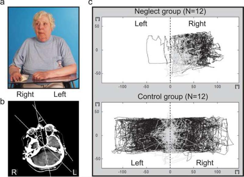
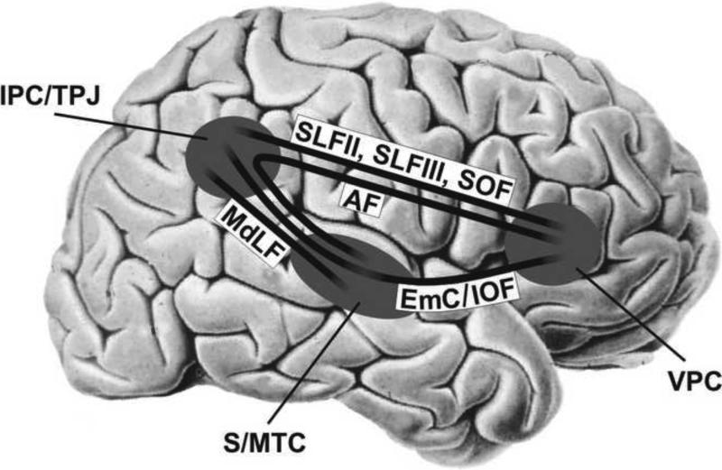
Spatial neglect is often perceived as a "heterogeneous collection of symptoms" with controversial anatomical correlates. However, a clear framework for core and satellite symptoms exists. Here we review the literature when viewed from the perspective of these different syndromes, and find clear pattern of anatomical injury. Specifically, the combined symptoms of biased gaze direction and search - with no awareness of these symptoms-is seen following structural damage to (particularly right hemisphere) perisylvian regions. Object centered deficits such as biased line bisection are due to more posterior (and possibly inferior) injury. Finally, extinction is associated with damage to the temporo-parietal junction. Further, we describe key choices that must be made to parse the spatial and attentional syndromes that result from right hemisphere injury, including the investigation of both acute and chronic injury as well as the use of functional and structural modalities.


Figures

Figure 1
Figure 1
(A) Example of the spontaneous eye and head orientation of patients with spatial neglect following a right hemispheric stroke while “doing nothing”, i.e. just sitting and waiting. The patients typically orient eyes and head towards the ipsilesional, right side. One could have the impression that the patient was fixating a certain target situated on the right side. However, the room was empty with only the photographer standing right in front of her. (Modified from Fruhmann-Berger & Karnath, 2005). (B) The eye-in-head deviation is even evident on the clinical brain scans taken at admission where the participant is simply asked to remain still. The egocentric bias is specifically associated with spatial neglect rather than with brain damage per se. (Modified from Becker & Karnath, 2010). (C) Scan paths (=gaze [combined eye and head orientation]) of a group of 12 patients with spatial neglect (upper panel) during active visual search (black lines) as well as at rest (gray lines) as compared with a group of 12 control patients without neglect (lower panel). The neglect patients show a marked bias of their active and their passive behavior toward the ipsilesional, right side. (Modified from Fruhmann-Berger et al., 2008.)
Figure 2
Figure 2
Sketch of the perisylvian neural network linking the inferior parietal lobule with the ventrolateral frontal cortex (via SLF II, SLF III, SOF), ventrolateral frontal cortex with superior/middle temporal cortex and insula (via AF, EmC/IOF), and superior temporal cortex with the inferior parietal lobule (via MdLF, EmC/IOF). SLF II/III, subcomponents II/III of the superior longitudinal fasciculus; SOF, superior occipitofrontal fasciculus; AF, arcuate fasciculus; EmC, extreme capsule; IOF, inferior occipitofrontal fasciculus; MdLF, middle longitudinal fasciculus; IPC, inferior parietal cortex; TPJ, temporo-parietal junction; S/MTC, superior/middle temporal cortex; VPC, ventrolateral prefrontal cortex. (Modified from Karnath, 2009).
Figure 3
Figure 3
(A) FLAIR images of the same patient in the acute and the chronic phase of a stroke lesion. Secondary lesion shrinkage is accompanied by sulcal widening and leads to diminished size of the chronic lesion. (B) Lesion ROI of a chronic periventricular lesion demarcated on a clinical FLAIR image before (I) and after SPM normalization (II). The same normalized ROI projected onto the standard template of a healthy brain (Ch2 single subject brain distributed with MRIcron) (III) results in a more lateral localization and erroneously indicates that the corpus of the caudate nucleus was “intact”, although it was actually lesioned in this individual (cf. I).
Figure 4
Figure 4
Statistical voxelwise lesion-behavior mapping (VLBM) analyses of 54 right hemisphere damaged patients based on their neglect severity scores measured in the acute and in the chronic phase of the stroke. Injury to highlighted regions predicts acute (upper panel) and chronic (lower panel) spatial neglect. Lesion of the superior and middle temporal cortex predicted both acute as well as chronic neglect. At the subcortical level the basal ganglia as well as the inferior occipitofrontal fasciculus/extreme capsule appeared to play a significant role for both acute as well as chronic neglect. MNI coordinates of each transverse section are given. (Modified from Karnath et al., 2011)

References

    1. Altamura C, Reinhard M, Vry MS, Kaller CP, Hamzei F, Vernieri F, Rossini PM, Hetzel A, Weiller C, Saur D. The longitudinal changes of BOLD response and cerebral hemodynamics from acute to subacute stroke. A fMRI and TCD study. BMC Neuroscience. 2009;10:151. - PMC - PubMed
    1. Becker E, Karnath H-O. Incidence of visual extinction after left versus right hemisphere stroke. Stroke. 2007;38:3172–3174. - PubMed
    1. Becker E, Karnath H-O. Neuroimaging of eye position reveals spatial neglect. Brain. 2010;133:909–914. - PubMed
    1. Behrmann M, Watt S, Black SE, Barton JJ. Impaired visual search in patients with unilateral neglect: an oculographic analysis. Neuropsychologia. 1997;35:1445–1458. - PubMed
    1. Bense S, Stephan T, Yousry TA, Brandt T, Dieterich M. Multisensory cortical signal increases and decreases during vestibular galvanic stimulation (fMRI). Journal of Neurophysiology. 2001;85:886–899. - PubMed

Publication types