. 2011 Sep 15;200(2):121-8.
doi: 10.1016/j.jneumeth.2011.06.020. Epub 2011 Jul 2.

A novel method for producing mono-biotinylated, biologically active neurotrophic factors: an essential reagent for single molecule study of axonal transport

Affiliations

A novel method for producing mono-biotinylated, biologically active neurotrophic factors: an essential reagent for single molecule study of axonal transport

Kijung Sung et al. J Neurosci Methods. .

Abstract

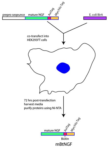
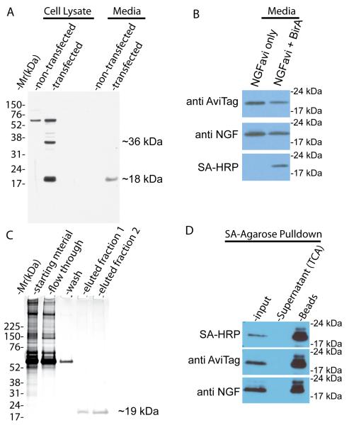
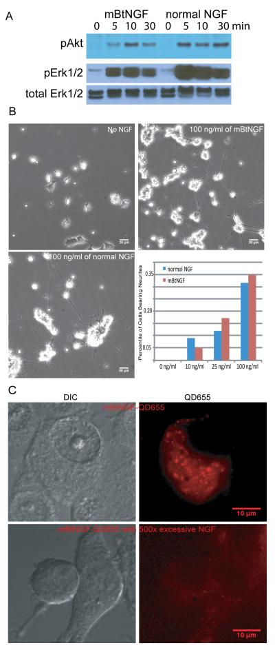
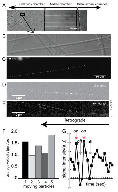
In this report, we describe a novel method for producing mature and biologically active mono-biotinylated nerve growth factors (mBtNGF) that can be used for single molecule studies of real-time movement of neurotrophins within axons of neurons. We inserted an AviTag sequence into the C-terminal of the full length mouse preproNGF cDNA and cloned the fusion construct into a pcDNA3.1 mammalian expression vector. We also subcloned the Escherichia coli biotin ligase, BirA, into a pcDNA3.1 vector. These two plasmids were then transiently co-expressed in HEK293FT cells. As a result, the AviTag located in the C-terminal of preproNGF was selectively ligated to a single biotin by BirA. The prepro sequence of NGF was subsequently cleaved within the cell. Mature mono-biotinylated NGF (mBtNGF) was secreted into cell culture media and was purified using Ni resin. We carried out activity assays and our results showed that mBtNGF retained biological activities that were comparable to normal NGF purified from mouse sub maxillary glands. We further verified the biotinylation efficiency of mBtNGF and the level of non-biotinylated NGF was virtually undetectable in the final preparation. Finally, by conjugating to quantum-dot streptavidin, mBtNGF was successfully used for single molecule study of axonal NGF trafficking in neurons.


Figures

Figure 1
Figure 1
Experimental designs for cloning, expression and purification of mBtNGF. As described in Materials and Methods, a mouse cDNA sequence encoding the full length of prepro NGF with a C-terminal AviTag is co-transfected with an expression vector for the E.coli biotin ligase (BirA) into 293FT cells. The specific “Lysine” residue within the AviTag is ligated to a biotin by BirA. Upon or during secretion, the N-terminal prepro sequence is cleaved and the mature protein with the biotinylated AviTag is released into the media. Mono-biotinylated NGF is secreted into the media and can be recovered from the media by Ni-NTA purification.
Figure 2
Figure 2
Purification and biochemical characterization of mature mBtNGF. A: Detection of the mature form, but not the immature form, of mBtNGF in the culture media of 293FT cells following transfection. Cell lysates and media collected from transfected and non-transfected 293FT cells were separated by SDS-PAGE and blotted with anti-AviTag antibodies. The unprocessed full-length protein (~35 kDa) and the mature protein (~19 kDa) are marked. B: BirA-dependent biotinylation. Media from cells that were double-transfected with the NGFavi construct and the BirA construct (NGF + BirA) or with the NGFavi construct alone (NGF only) were separated by SDS-PAGE and blotted with indicated antibodies. The biotinylated forms were detected only with streptavidin-HRP (SA-HRP). C: Purification of mature mBtNGF to apparent homogeneity. Starting materials, flow-through, wash and two eluted fractions (25 μl each) from Ni-NTA column purification were separated by SDS-PAGE followed by silver staining. The eluted fractions contains only the ~19kDa species that corresponds to the mature mBtNGF. D: Assay for biotinylation efficiency of mBtNGF. Purified mBtNGF was incubated with streptavidin-agarose beads for 2hr at 4°C. The beads were washed and boiled in SDS sample buffer. The supernatants were collected and precipitated with TCA. These samples along with input were analyzed by SDS-PAGE and immunoblotting. The blot was probed with SA-HRP, with anti AviTag and anti NGF antibodies. All signals were detected in the SA-bead sample and no signal was seen in the supernatant.
Figure 3
Figure 3
Analysis of bioactivities of mBtNGF using PC12 cells. A: Activation of Akt and Erk1/2 signaling pathways by mBtNGF. PC12 cells were serum-starved and were stimulated for 0, 5, 10 and 30 min with either 100ng/ml mBtNGF or 100 ng/ml normal NGF. Cell lysates were analyzed by SDS-PAGE/immunoblotting with indicated antibodies. B: Neurite outgrowth assay. PC12 cells were treated with vehicle only (0), or 10, 25 or 100ng/ml mBtNGF or normal NGF at the same concentrations for 6 days. The percentage of cells bearing neurites longer than three times of cell body was presented. Typical micrographs of PC12 cells treated with the vehicle (no NGF), 100 ng/ml mBtNGF or normal NGF were shown. C: Assay for internalization of mBtNGF. PC12 cells after incubating with either 0.2 nM of mBtNGF-QD655 or with 0.2 nM mBtNGF-QD655 together with 100 nM of normal NGF. Both DIC and corresponding fluorescence images were captured and shown.
Figure 4
Figure 4
Single molecule imaging of transport of mBtNGF-QD655 in axons of dorsal root ganglion (DRG). A: A phase contrast image of DRG neurons that were cultured in the microfluidic chambers. Following addition of 0.2 nM mBtNGF-QD655 to the distal axons, real time imaging of transport of mBtNGF-QD655 within the proximal axons was carried out using a 100x oil objective lens. A high resolution DIC image of proximal axons of DRG neurons is shown in B. Time-lapse trajectory of mBtNGF-QD655 movement within the corresponding axons is shown in C. Kymograph analysis of Movie 2 (supplemental materials) was also performed using ImageJ. The result of Z-project is shown in D and the resulting kymograph is shown in E. F: A histogram shows the average velocities of retrogradely transported endosomes. G: A line graph shows the blinking properties of single QDs. Fluorescence intensity of a moving particle was measured frame by frame. Following background subtraction, the signal intensity displays a pattern of either complete “on” or complete “off”, characteristic of blinking of a single QD, whereas an out-of-focus signal should show a slow drop in fluorescence intensity instead.

Similar articles

Cited by

References

    1. Adachi N, Kohara K, Tsumoto T. Difference in trafficking of brain-derived neurotrophic factor between axons and dendrites of cortical neurons, revealed by live-cell imaging. BMC Neurosci. 2005;6:42. - PMC - PubMed
    1. Beckett D, Kovaleva E, Schatz PJ. A minimal peptide substrate in biotin holoenzyme synthetase-catalyzed biotinylation. Protein Sci. 1999;8:921–9. - PMC - PubMed
    1. Bronfman FC, Tcherpakov M, Jovin TM, Fainzilber M. Ligand-induced internalization of the p75 neurotrophin receptor: a slow route to the signaling endosome. J Neurosci. 2003;23:3209–20. - PMC - PubMed
    1. Chao MV. Neurotrophins and their receptors: a convergence point for many signalling pathways. Nat Rev Neurosci. 2003;4:299–309. - PubMed
    1. Chao MV, Rajagopal R, Lee FS. Neurotrophin signalling in health and disease. Clin Sci (Lond) 2006;110:167–73. - PubMed

Publication types

MeSH terms