

Eicosanoids, β-cell function, and diabetes
- PMID: 21757024
- PMCID: PMC3144311
- DOI: 10.1016/j.prostaglandins.2011.06.001
Eicosanoids, β-cell function, and diabetes
Abstract
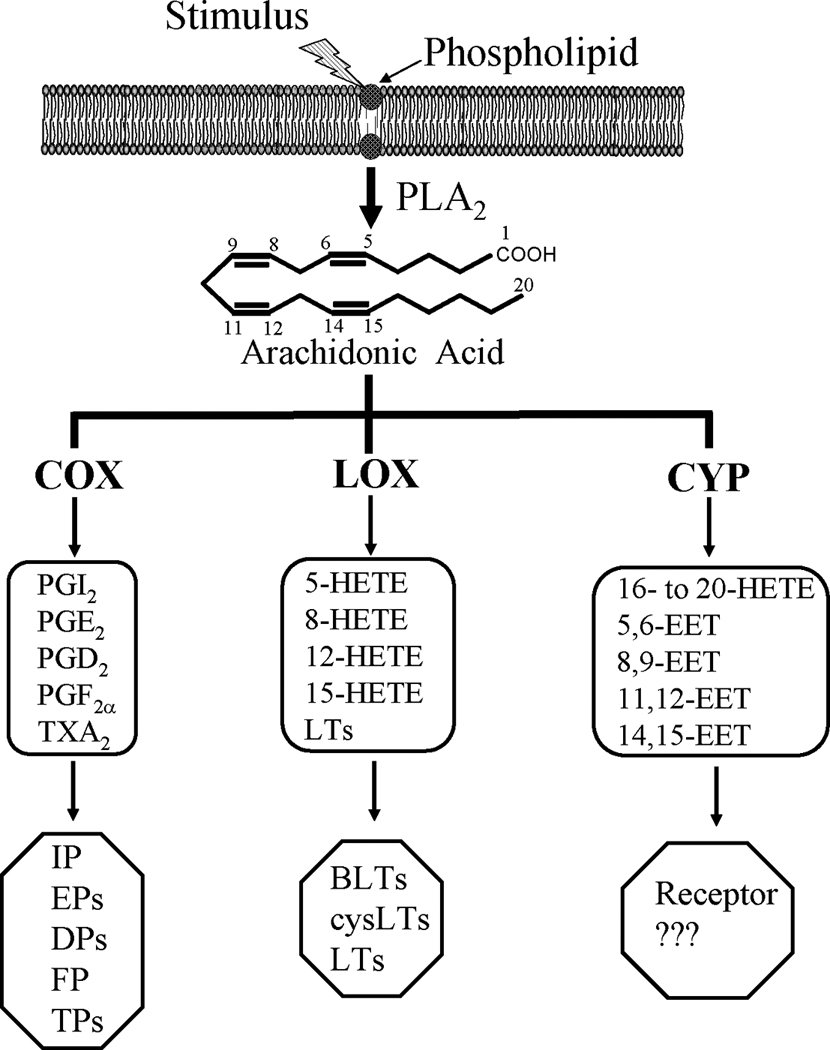
Arachidonic acid (AA) is metabolized by cyclooxygenase (COX), lipoxygenase (LOX), and cytochrome P450 (CYP) enzymes into eicosanoids, which are involved in diverse diseases, including type 1 and type 2 diabetes. During the last 30 years, evidence has been accumulated that suggests important functions for eicosanoids in the control of pancreatic β-cell function and destruction. AA metabolites of the COX pathway, especially prostaglandin E(2) (PGE(2)), appear to be significant factors to β-cell dysfunction and destruction, participating in the pathogenesis of diabetes and its complications. Several elegant studies have contributed to the sorting out of the importance of 12-LOX eicosanoids in cytokine-mediated inflammation in pancreatic β cells. The role of CYP eicosanoids in diabetes is yet to be explored. A recent publication has demonstrated that stabilizing the levels of epoxyeicosatrienoic acids (EETs), CYP eicosanoids, by inhibiting or deleting soluble epoxide hydrolase (sEH) improves β-cell function and reduces β-cell apoptosis in diabetes. In this review we summarize recent findings implicating these eicosanoid pathways in diabetes and its complications. We also discuss the development of animal models with targeted gene deletion and specific enzymatic inhibitors in each pathway to identify potential targets for the treatment of diabetes and its complications.
Copyright © 2011 Elsevier Inc. All rights reserved.
Figures
References
-
- Shaw JE, Sicree RA, Zimmet PZ. Global estimates of the prevalence of diabetes for 2010 and 2030. Diabetes Res Clin Pract. 2010;87:4–14. - PubMed
-
- Yoon JW, Jun HS. Autoimmune destruction of pancreatic beta cells. Am J Ther. 2005;12:580–591. - PubMed
-
- Cnop M, Welsh N, Jonas JC, et al. Mechanisms of pancreatic beta-cell death in type 1 and type 2 diabetes: many differences, few similarities. Diabetes. 2005;54:S97–S107. - PubMed
-
- Eizirik DL, Colli ML, Ortis F. The role of inflammation in insulitis and beta-cell loss in type 1 diabetes. Nat Rev Endocrinol. 2009;5:219–226. - PubMed
Publication types
MeSH terms
Substances
Grants and funding
LinkOut - more resources
Full Text Sources
Medical
