. 2011 Jul 15;71(14):4834-45.
doi: 10.1158/0008-5472.CAN-11-0027. Epub 2011 Jul 14.

Human neural stem cell transplantation ameliorates radiation-induced cognitive dysfunction

Affiliations

Human neural stem cell transplantation ameliorates radiation-induced cognitive dysfunction

Munjal M Acharya et al. Cancer Res. .

Abstract

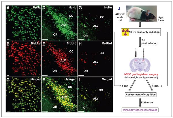
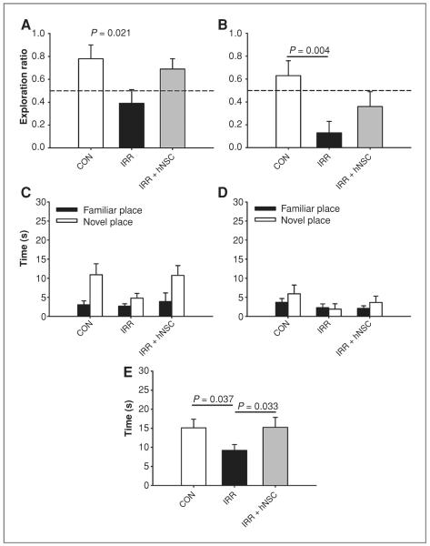
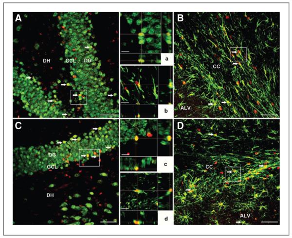
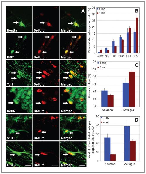
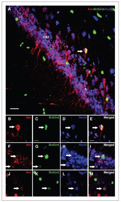
Cranial radiotherapy induces progressive and debilitating declines in cognition that may, in part, be caused by the depletion of neural stem cells. The potential of using stem cell replacement as a strategy to combat radiation-induced cognitive decline was addressed by irradiating athymic nude rats followed 2 days later by intrahippocampal transplantation with human neural stem cells (hNSC). Measures of cognitive performance, hNSC survival, and phenotypic fate were assessed at 1 and 4 months after irradiation. Irradiated animals engrafted with hNSCs showed significantly less decline in cognitive function than irradiated, sham-engrafted animals and acted indistinguishably from unirradiated controls. Unbiased stereology revealed that 23% and 12% of the engrafted cells survived 1 and 4 months after transplantation, respectively. Engrafted cells migrated extensively, differentiated along glial and neuronal lineages, and expressed the activity-regulated cytoskeleton-associated protein (Arc), suggesting their capability to functionally integrate into the hippocampus. These data show that hNSCs afford a promising strategy for functionally restoring cognition in irradiated animals.


Figures

Figure 1
Figure 1
In vitro and in vivo colabeling of HuNu (green) and BrdUrd (red). Dual labeling of HuNu with BrdUrd in vitro (A–C) and in vivo at 1 month (D–F) and 4 month (G–I) postgrafting in host hippocampus. ALV, alves of hippocampus; OR, oriens of hippocampus. Schematic illustration of transplantation studies (J). Two-month-old athymic nude rats receiving 10 Gy head-only irradiation were engrafted with hNSCs 2 days later. At 1 or 4 months following surgery, animals were subjected to cognitive testing, using the hippocampal dependent NPR task and then euthanized for engrafted cell survival and differentiation analyses. Control and irradiated animals receiving sterile vehicle served as sham surgery groups. Scale bars: A–C, 10 μm; D–I, 50 μm.
Figure 2
Figure 2
Human neural stem cell engraftment improves radiation-induced impairments in NPR assessed 1 month postimplantation. A and B, exploration ratios (i.e., tnovel/tnovel + tfamiliar) for the first minute of the 5-minute and 24-hour test sessions, respectively. C and D, plots of total exploration time over the entire 3-minute test session for the 5-minute and 24-hour test sessions, respectively. E, time spent exploring both objects during the initial familiarization phase. Data are presented as means + SEM, and the dashed lines (in A and B) indicate chance performance (i.e., 0.5). P values are derived from FPLSD post hoc comparisons.
Figure 3
Figure 3
Human neural stem cell engraftment improves radiation-induced impairments in NPR assessed 4 months postimplantation. A and B, exploration ratios (i.e., tnovel/tnovel + tfamiliar) for the first minute of the 5-minute and 24-hour test sessions, respectively. C and D, plots of total exploration time over the entire 3-minute test sessions for the 5-minute and 24-hour test sessions, respectively. E, time spent exploring both objects during the initial familiarization phase. Data are presented as means + SEM, and the dashed lines (in A and B) indicate chance performance. P values are derived from FPLSD post hoc comparisons.
Figure 4
Figure 4
Engrafted hNSCs survive and migrate in the host hippocampus. At 1 month postengraftment, hNSCs showed extensive migration from the injection site throughout the hippocampus [DG, DH, granule cell layer (GCL), CA1 and CA3 subfields, and partially in the CC; magnification ×4–40 (in A–D)]. Grafted cells were detected by BrdUrd immunostaining (dark brown nuclei, indicated by red arrows; D) and counterstained with hematoxylin. Images were derived from irradiated animals engrafted with hNSCs and analyzed at 1 month postgrafting. Enumeration of transplanted hNSCs by unbiased stereology revealed that 23% and 12% of the engrafted cells survived at 1- and 4-month postgrafting time points, respectively (E). Scale bars: A, 200 μm; B, 100 μm; C, 50 μm; D, 20 μm. ***, P = 0.043 comparing 1-month and 4-month postengraftment groups.
Figure 5
Figure 5
Differentiation of transplanted hNSCs in the irradiated hippocampus. At 1 month postgrafting (A, B and a, b), BrdUrd-positive (red) hNSCs differentiated into NeuN-positive (green) mature neurons (A and a) and GFAP-positive (green) astrocytes(Bandb)as visualizedby dual labeling of neuron- or astrocyte-specific markers. Similar phenotypes were observed at 4 months postgrafting (C, D and c, d), where BrdUrd-positive (red) engrafted cells were colabeled with markers of mature neurons (NeuN, green; C and c), and astrocytes (GFAP, green; D and d). Arrows indicate dual labeled grafted cells, and boxes represent regions magnified for orthogonal Z-stacks (in A–D). Orthogonal reconstructions of BrdUrd+/NeuN+ colabeled cells (a and c) and BrdUrd+/GFAP+ colabeled cells (b and d) are shown at each postgrafting time point. GCL, granule cell layer; ALV, alves of hippocampus; DG, dentate gyrus; DH, dentate hilus. Scale bars: A–D, 50 μm; a–d, 10 μm.
Figure 6
Figure 6
Transplanted hNSCs differentiated into neuronal and astrocytic phenotypes at 1 and 4 months postengraftment. A and B, hNSCs positive for BrdUrd occasionally expressed the cell-cycle marker (Ki67) and multipotent marker (nestin). Engrafted (BrdUrd+) cells differentiated into immature (Tuj1+) and mature (NeuN+) neurons (A and B). The majority of engrafted (BrdUrd+) cells differentiated into mature (S100+, S100ß protein) and immature (GFAP+) astrocytes (A and B). Arrows indicate dual-labeled engrafted cells. The percentage (C) and yield (D) of differentiated hNSCs within each hemisphere of the irradiated rat brain. Scale bars: A, 5 μm.
Figure 7
Figure 7
HNSC graft–derived cells express behaviorally induced Arc in the irradiated hippocampus. Irradiated animals engrafted with hNSCs and subjected to a hippocampal-dependent novelty exploration task at 1 month postgrafting were rapidly processed for the isolation and analysis of brain tissues. Engrafted hNSCs (BrdUrd + cells, green; A, C, G, and K) + differentiated into NeuN-positive mature neurons (blue; A, D, H, and L) and expressed Arc (red; A, B, F, and J) as visualized by the triple labeling of neuron- and Arc-specific markers (A, E, I, and M). Arrows indicate triple-labeled cells. Scale bars: A, 50 μm; B–M, 10 μm.

References

    1. Abayomi OK. Pathogenesis of irradiation-induced cognitive dysfunction. Acta Oncol. 1996;35:659–63. - PubMed
    1. Anderson VA, Godber T, Smibert E, Weiskop S, Ekert H. Cognitive and academic outcome following cranial irradiation and chemotherapy in children: a longitudinal study. Br J Cancer. 2000;82:255–62. - PMC - PubMed
    1. Raber J. Unintended effects of cranial irradiation on cognitive function. Toxicol Pathol. 2010;38:198–202. - PubMed
    1. Meyers CA, Geara F, Wong PF, Morrison WH. Neurocognitive effects of therapeutic irradiation for base of skull tumors. Int J Radiat Oncol Biol Phys. 2000;46:51–5. - PubMed
    1. Roman DD, Sperduto PW. Neuropsychological effects of cranial radiation: current knowledge and future directions. Int J Radiat Oncol Biol Phys. 1995;31:983–98. - PubMed

Publication types

Substances