

Low-dose temporal bone CT in infants and young children: effective dose and image quality
- PMID: 21757514
- PMCID: PMC7964352
- DOI: 10.3174/ajnr.A2524
Low-dose temporal bone CT in infants and young children: effective dose and image quality
Abstract
Background and purpose: The temporal bone is ideal for low-dose CT because of its intrinsic high contrast. The aim of this study was to retrospectively evaluate image quality and radiation doses of a new low-dose versus a standard high-dose pediatric temporal bone CT protocol and to review dosimetric data from the literature.
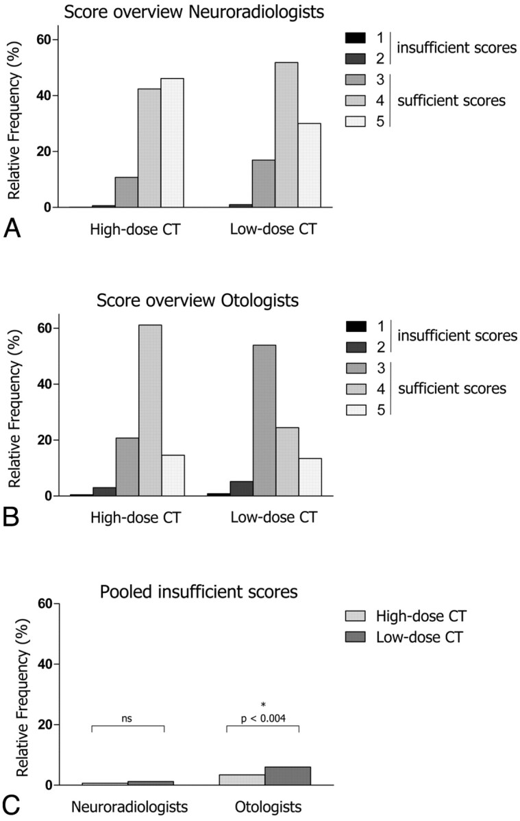
Materials and methods: Image quality and radiation doses were compared for 38 low-dose (80 kV/90-110 mAs) and 16 high-dose (140 kV/170 mAs) temporal bone CT scans of infants to 5-year-old children. The CT visualization quality of 23 middle and inner ear structures was subjectively graded by 3 neuroradiologists and 3 otologists by using a 5-point scale with scores 1-2 indicating insufficient and scores 3-5 indicating sufficient image quality. Effective doses of local and literature-derived protocols were calculated from dosimetric data by using NRPB-SR250 software.
Results: Insufficient image-quality scores were more frequent in low-dose scans versus high-dose scans, but the difference was only statistically significant for otologists (6.0% versus 3.4%, P = .004) and not for neuroradiologists (1.2% versus 0.7%, P = .84). Image quality was critical for small structures (such as the stapes or lamella at the internal auditory canal fundus). Effective doses were 0.25-0.3 mSv for low-dose scans, 1.4-1.8 mSv for high-dose scans, and 0.9-2.6 mSv for literature-derived protocols.
Conclusions: The image quality of the new low-dose protocol remains diagnostic for assessing middle and inner ear anatomy despite a 3- to 8-fold dose reduction over previous and literature-derived protocols. However, image quality of small structures is critical and may be perceived as insufficient.
Figures
References
-
- Radiation Risks and Pediatric Computed Tomography (CT): A Guide for Health Care Providers. Rockville, Maryland: National Cancer Institute. 2008. Available at http//:www.cancer.gov/cancertopics/causes/radiation-risks-pediatric-CT. Accessed December 10, 2010
-
- Vock P. CT dose reduction in children. Eur Radiol 2005;15:2330–40 - PubMed
-
- Tack D. Methods and strategies for radiation dose optimization–and reduction–in MDCT with special focus on the image quality in computed tomography. In: Baert AL, Knauth M, Sartor K. Radiation Dose from Adult and Pediatric Multidetector CT. Berlin: Springer-Verlag; 2007:112
-
- Husstedt HW, Prokop M, Dietrich B, et al. Low-dose high-resolution CT of the petrous bone. J Neuroradiol 2000;27:87–92 - PubMed
-
- Lutz J, Jäger V, Hempel MJ, et al. Delineation of temporal bone anatomy: feasibility of low-dose 64-row CT in regard to image quality. Eur Radiol 2007;17:2638–45 - PubMed
MeSH terms
LinkOut - more resources
Full Text Sources
Medical