Case Reports
. 2011 Oct;32(9):1703-6.
doi: 10.3174/ajnr.A2569. Epub 2011 Jul 14.

Oncocytoma: the vanishing parotid mass

Affiliations
Case Reports

Oncocytoma: the vanishing parotid mass

N D Patel et al. AJNR Am J Neuroradiol. 2011 Oct.

Abstract

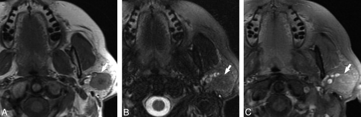
Parotid gland oncocytoma is an uncommon, benign salivary neoplasm composed of mitochondria-rich oncocytes. The purpose of this study was to correlate MR imaging and histopathology of parotid gland oncocytomas and to define the features that may distinguish these neoplasms from other benign and malignant parotid gland tumors. The MR imaging features in 9 patients with a pathologic diagnosis of oncocytoma were retrospectively reviewed. The imaging features were strikingly similar for 8 of the 9 patients. All lesions appeared T1 hypointense but isointense to the native parotid gland on fat-saturated T2 and postcontrast T1 imaging. On MR imaging, parotid gland oncocytomas share specific imaging characteristics that have not been described for benign or malignant parotid gland tumors. Oncocytomas are isointense to native parotid gland on fat-saturated T2 and T1 postcontrast MR images. Preoperative identification of correct histology may help surgical planning.


Figures

Fig 1.
Fig 1.
Case 1, 62-year-old female with 2-year history of firm, painless left parotid mass. A, Axial T1-weighted MR image shows a well-demarcated mass within the superficial lobe of the left parotid gland (white arrow) that is T1 hypointense. B, Axial T2-weighted MR image with fat saturation shows the mass (white arrow) to be isointense to the native parotid gland. C, Axial T1-weighted postgadolinium sequences illustrates the mass (white arrow) as isointense to the native parotid gland.
Fig 2.
Fig 2.
Case 5, 55-year-old woman with painless “lump” behind her left ear. A, Axial T1-weighted MR image shows a well-demarcated mass within the superficial and deep lobe of the left parotid gland (white arrow) that is T1 hypointense. B, Axial T2-weighted MR image with fat saturation illustrates the mass (white arrow) to be isointense to the native parotid gland. C, Axial T1-weighted postgadolinium sequence shows the mass (white arrow) as isointense to the native parotid gland.
Fig 3.
Fig 3.
Case 7, 75-year-old man with history of squamous cell carcinoma of the larynx status postradiation found to have an enlarging mass in the left parotid gland on surveillance imaging. A, Axial T1-weighted MR image shows a well-demarcated mass within the superficial lobe of the left parotid gland (white arrow) that is T1 hypointense. B, Axial T2-weighted MR image with fat saturation shows the mass to be hyperintense but heterogeneous to the native parotid gland. C, Axial T1-weighted postgadolinium sequence shows the mass (white arrow) to demonstrate heterogeneous enhancement to the native parotid gland.
Fig 4.
Fig 4.
Typical histologic features of a parotid gland oncocytoma including eosinophilic cells with finely granular cytoplasm and uniform, round, centrally placed nuclei (hematoxylin-eosin stain, original magnification, ×400).

References

    1. Huvos AG.Oncocytoma. In: Barnes L, Eveson JW, Reichart P, et al., eds. World Health Organization Classification of Tumors: Pathology and Genetics, Head and Neck Tumors. Lyon, France: IARC Press; 2005: 242–43
    1. Brandwein MS, Huvos AG. Oncocytic tumors of major salivary glands: a study of 68 cases with follow-up of 44 patients. Am J Surg Pathol 1991; 15: 514–28 - PubMed
    1. Shah VN, Branstetter BF. Oncocytoma of the parotid gland: a potential false-positive finding on 18F-FDG PET. AJR Am J Roentgenol 2007; 189: W212–14 - PubMed
    1. Branstetter BF, IV, Blodgett TM, Zimmer LA, et al. Head and neck malignancy: is PET/CT more accurate than PET or CT alone? Radiology 2005; 235: 580–86 - PubMed
    1. Tan TJ, Tan TY. CT features of parotid gland oncocytomas: a study of 10 cases and literature review AJNR Am J Neuroradiol 2010; 31: 1413–17 - PMC - PubMed

Publication types