

Variations in the course of the cervical vagus nerve on thyroid ultrasonography
- PMID: 21757523
- PMCID: PMC7966036
- DOI: 10.3174/ajnr.A2476
Variations in the course of the cervical vagus nerve on thyroid ultrasonography
Abstract
Background and purpose: Only 1 ultrasonography study that described the variation of the VN had been published at the time our research was begun. The purpose of this study was to evaluate the incidence and type of variation in the course of the cervical VN on thyroid ultrasonography.
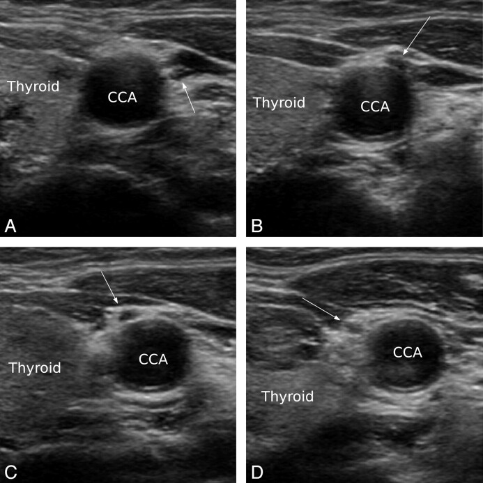
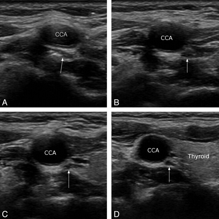
Materials and methods: From August 2009 to September 2010, 163 consecutive patients were evaluated by sonography for the screening and characterization of thyroid nodules (mean age, 49.0 ± 14.4 years, male:female, 20:143). Two types of variation were defined as follows: 1) anterior variation, when the course of the VNs changed from the typical location to an anterior location in front of the CCA; and 2) medial variation, when the course of the VNs changed from the typical location to a location medial to the CCA (between CCA and thyroid gland). The incidence of the each variation was studied.
Results: Variation in the course of the VN occurred in 5.5% (18/326) of cases. The anterior variation was observed in 4.3% (14/326, right:left = 4:10), and the medial variation was observed in 1.2% (4/326, right:left = 3:1). For both variations, the VN was close to or nearly abutted the thyroid gland after it changed course.
Conclusions: Variation in the course of the cervical VN could be assessed by ultrasonography. Two variations were observed in 5.5% of cases. The anterior variation was more common than the medial variation.
Figures

References
-
- Knappertz VA, Tegeler CH, Hardin SJ, et al. Vagus nerve imaging with ultrasound: anatomic and in vivo validation. Otolaryngol Head Neck Surg 1998;118:82–85 - PubMed
-
- Giovagnorio F, Martinoli C. Sonography of the cervical vagus nerve: normal appearance and abnormal findings. AJR Am J Roentgenol 2001;176:745–49 - PubMed
-
- AbuRahma AF, Lim RY. Management of vagus nerve injury afer carotid endarterectomy. Surgery 1996;119:245–47 - PubMed
-
- Ballotta E, Da Giau G, Renon L, et al. Cranial and cervical nerve injuries after carotid endarterectomy: a prospective study. Surgery 1999;125:85–91 - PubMed
Publication types
MeSH terms
LinkOut - more resources
Full Text Sources