. 2011 Sep;22(17):3206-17.
doi: 10.1091/mbc.E11-02-0145. Epub 2011 Jul 14.

Phosphorylation of Serine 114 on Atg32 mediates mitophagy

Affiliations

Phosphorylation of Serine 114 on Atg32 mediates mitophagy

Yoshimasa Aoki et al. Mol Biol Cell. 2011 Sep.

Abstract

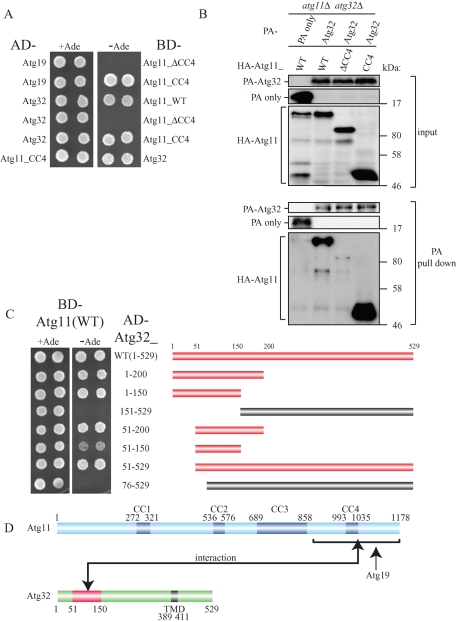
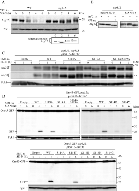
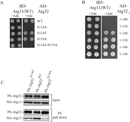
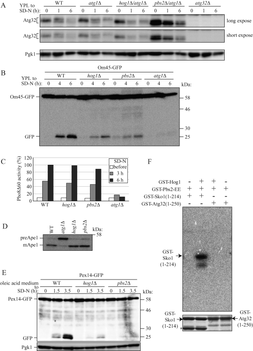
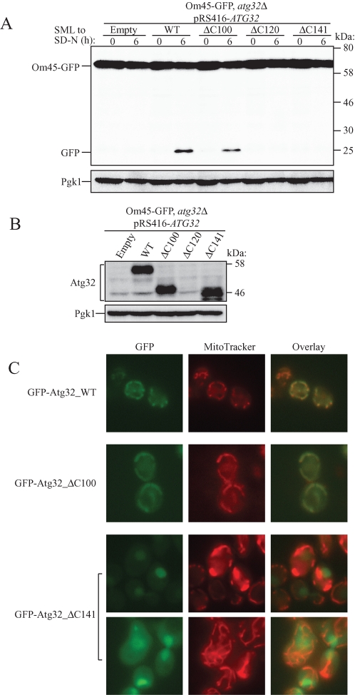
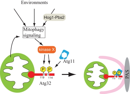
Mitophagy, which selectively degrades mitochondria via autophagy, has a significant role in mitochondrial quality control. When mitophagy is induced in yeast, mitochondrial residential protein Atg32 binds Atg11, an adaptor protein for selective types of autophagy, and it is recruited into the vacuole along with mitochondria. The Atg11-Atg32 interaction is believed to be the initial molecular step in which the autophagic machinery recognizes mitochondria as a cargo, although how this interaction is mediated is poorly understood. Therefore, we studied the Atg11-Atg32 interaction in detail. We found that the C-terminus region of Atg11, which included the fourth coiled-coil domain, interacted with the N-terminus region of Atg32 (residues 100-120). When mitophagy was induced, Ser-114 and Ser-119 on Atg32 were phosphorylated, and then the phosphorylation of Atg32, especially phosphorylation of Ser-114 on Atg32, mediated the Atg11-Atg32 interaction and mitophagy. These findings suggest that cells can regulate the amount of mitochondria, or select specific mitochondria (damaged or aged) that are degraded by mitophagy, by controlling the activity and/or localization of the kinase that phosphorylates Atg32. We also found that Hog1 and Pbs2, which are involved in the osmoregulatory signal transduction cascade, are related to Atg32 phosphorylation and mitophagy.


Figures

FIGURE 1:
FIGURE 1:
The N-terminus region (51–150) of Atg32 interacts with the C-terminus region of Atg11. (A) Yeast two-hybrid analysis between Atg19 and Atg11 mutants, and between Atg32 and Atg11 mutants. The PJ69-4A strain was transformed with pGAD and pGBD plasmid, which can express the indicated proteins. Cells were grown on +Ade or –Ade plates at 30°C for 4 d. (B) The C-terminus region of Atg11 associated with Atg32 during nitrogen starvation. The atg11Δ atg32Δ strains expressing PA-tagged Atg32 or PA only and the indicated HA-tagged Atg11 mutants under the control of the CUP1 promoter were grown in SMD medium until the mid–log phase and then starved in SD-N for 1 h. PA-Atg32 was precipitated using IgG–Sepharose from cell lysates. Top two, an immunoblot of total-cell lysates. Bottom two, the IgG precipitates, which were probed with anti-HA and anti-PA antibodies. (C) Yeast two-hybrid analysis between Atg11 and the indicated Atg32 mutants (left). Each bar indicates expressed Atg32 mutants (right). Red bars indicate yeast two-hybrid positive and the gray bars indicate yeast two-hybrid negative. (D) Schematic drawing of Atg11 and Atg32. Atg11 is predicted to have a fourth coiled-coil domain (CC1–CC4). Atg32 has a TMD. The domain of Atg32 required for Atg11 interaction as determined by yeast two-hybrid study is highlighted in red.
FIGURE 2:
FIGURE 2:
Phosphorylation of Atg32, especially phosphorylation of Ser-114 on Atg32, is essential for mitophagy. (A) WT or atg11Δ strains were cultured in YPL medium until the mid–log growth phase (indicated as b) and then shifted to SD-N medium for 0, 2, and 6 h. The amount and the modification of endogenous Atg32 were observed by immunoblotting with anti-Atg32 and anti-Por1 (loading control) antibodies. The variation in molecular weight of Atg32 in the atg11Δ strain is shown as a schematic model. The asterisk indicates a nonspecific band. (B) The atg11Δ strain was cultured in YPL medium until the mid–log growth phase (before SD-N) and then shifted to SD-N for 6 h. Cell lysates were treated with or without λ PPase at 30°C for 1 h. The molecular weight change was observed by immunoblotting with anti-Atg32 antibody. (C) The atg32Δ atg11Δ double-knockout strain transformed with vectors expressing the indicated ATG32 mutants were cultured in YPL medium until the mid–log growth phase and then shifted to SD-N medium for 0, 1, and 6 h. The modification of Atg32 mutants was monitored by immunoblotting with anti-Atg32 and anti-Pgk1 (loading control) antibodies. The asterisks indicate a long exposure. (D) Strains of atg32Δ expressing Om45-GFP were transformed with the indicated Atg32 mutant-expressing vectors. Cells were cultured in SML medium until the mid–log growth phase and then shifted to SD-N for 6 h. GFP processing was monitored by immunoblotting with anti-GFP and anti-Pgk1 (loading control) antibodies.
FIGURE 3:
FIGURE 3:
Phosphorylation of Ser-114 on Atg32 is critically important for the Atg11–Atg32 interaction. (A, B) Yeast two-hybrid analysis between Atg11 and the indicated Atg32 mutants. (C) The atg11Δ atg32Δ strains expressing PA-tagged Atg32WT, PA-tagged Atg32S114A, or PA-tagged Atg32S114T and HA-tagged Atg11 under the control of the CUP1 promoter were grown in SMD medium until the mid–log phase and then starved in SD-N for 1 h. PA-Atg32 was precipitated using IgG–Sepharose from cell lysates. Top two, an immunoblot of total-cell lysates. Bottom two, the IgG precipitates, which were probed with anti-HA and anti-PA antibodies.
FIGURE 4:
FIGURE 4:
Deletion of HOG1 or PBS2 affects both Atg32 phosphorylation and mitophagy. (A) WT, atg1Δ, and atg32Δ strains and hog1Δ/atg1Δ and pbs2Δ/atg1Δ double-knockout strains were cultured in YPL medium until the mid–log growth phase and then shifted to SD-N medium for 0, 1, and 6 h. Phosphorylation on endogenous Atg32 was monitored by immunoblotting with anti-Atg32 and anti-Pgk1 (loading control) antibodies. (B) Strains deleted for the indicated genes and expressing Om45-GFP were cultured in YPL medium until the mid–log growth phase and then shifted to SD-N for 4 and 6 h. GFP processing was monitored by immunoblotting with anti-GFP antibody. (C) The WT (TKMY236), hog1Δ (TKYM248), pbs2Δ (TKYM249), and atg1Δ (TKYM256) strains were grown in YPD medium and shifted to SD-N for 3 and 6 h. Samples were collected and protein extracts assayed for Pho8Δ60 activity. (D) WT, atg1Δ, hog1Δ, and pbs2Δ strains were cultured in YPD medium and analyzed for prApe1 maturation by immunoblotting with anti-Ape1 antiserum. (E) Strains deleted for the indicated genes and expressing Pex14-GFP were cultured with oleic acid–containing medium for 19 h and then shifted to SD-N for the indicated times and monitored for GFP processing by immunoblotting. (F) In vitro phosphorylation of Sko1(1-214) and Atg32(1-250) by Hog1. Recombinant GST-Sko1(1-214) or GST-Atg32(1-250) was phosphorylated by activated recombinant GST-Hog1 in the presence of [γ-32P]ATP. The labeled proteins were resolved by SDS–PAGE, and an autoradiograph image (top) and Coomassie Brilliant Blue–stained image (bottom) of the gel were taken. The asterisks indicate GST-Sko1 degradation product or nonspecific bands.
FIGURE 5:
FIGURE 5:
Characterization of the C-terminus region of Atg32. (A) Strains of atg32Δ expressing Om45-GFP were transformed with the indicated Atg32 mutant–expressing vectors. Cells were cultured in SML medium until the mid–log growth phase and then shifted to SD-N for 6 h. GFP processing was monitored by immunoblotting with anti-GFP and anti-Pgk1 (loading control) antibodies. (B) The same cells shown in A were cultured in SML medium until the mid–log growth phase. The expression level of Atg32 was observed by immunoblotting with anti-Atg32 and anti-Pgk1 (loading control) antibodies. (C) The wild-type strain transformed with a plasmid expressing the indicated GFP-tagged Atg32 mutants under the control of the CUP1 promoter was cultured in SMD medium until the mid–log growth phase. Cells were labeled with the mitochondrial marker MitoTracker Red and analyzed by fluorescence microscopy.
FIGURE 6:
FIGURE 6:
Model of mitophagy in yeast. The initial trigger-inducing mitophagy comes from mitochondria (such as mitochondrial damage) and/or the cellular environment (such as dramatic nutrient change and oxidative stresses). The initial trigger is through an unidentified mitophagy signaling pathway and it reaches kinase X, which is believed to be a Ser/Thr–specific protein kinase. This kinase phosphorylates Ser-114 and Ser-119 on Atg32. Phosphorylation of Atg32, especially phosphorylation of Ser-114, mediates the Atg11–Atg32 interaction. Atg11 recruits mitochondria to the PAS, where the autophagosome is generated to enclose the mitochondria. Hog1 and Pbs2 affect the mitophagy signaling pathway or affect kinase X.

References

    1. Arai M, Imai H, Koumura T, Yoshida M, Emoto K, Umeda M, Chiba N, Nakagawa Y. Mitochondrial phospholipid hydroperoxide glutathione peroxidase plays a major role in preventing oxidative injury to cells. J Biol Chem. 1999;274:4924–4933. - PubMed
    1. Bilsland-Marchesan E, Arino J, Saito H, Sunnerhagen P, Posas F. Rck2 kinase is a substrate for the osmotic stress-activated mitogen-activated protein kinase Hog1. Mol Cell Biol. 2000;20:3887–3895. - PMC - PubMed
    1. Bogenhagen DF. Repair of mtDNA in vertebrates. Am J Hum Genet. 1999;64:1276–1281. - PMC - PubMed
    1. Clotet J, Posas F. Control of cell cycle in response to osmostress: lessons from yeast. Methods Enzymol. 2007;428:63–76. - PubMed
    1. Cribbs JT, Strack S. Functional characterization of phosphorylation sites in dynamin-related protein 1. Methods Enzymol. 2009;457:231–253. - PMC - PubMed

Publication types

MeSH terms

Substances