. 2011 Sep;22(17):3041-54.
doi: 10.1091/mbc.E11-04-0321. Epub 2011 Jul 14.

Neuroprotective role of PrPC against kainate-induced epileptic seizures and cell death depends on the modulation of JNK3 activation by GluR6/7-PSD-95 binding

Affiliations

Neuroprotective role of PrPC against kainate-induced epileptic seizures and cell death depends on the modulation of JNK3 activation by GluR6/7-PSD-95 binding

Patricia Carulla et al. Mol Biol Cell. 2011 Sep.

Abstract

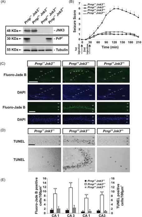
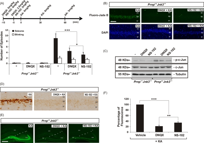
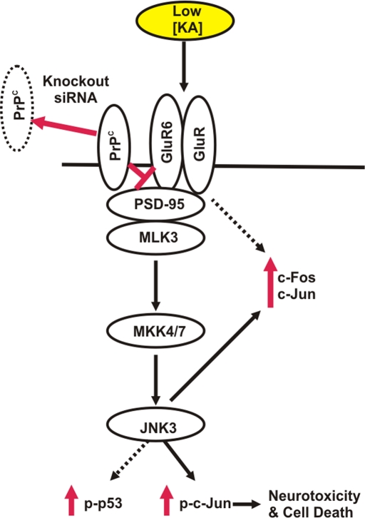
Cellular prion protein (PrP(C)) is a glycosyl-phosphatidylinositol-anchored glycoprotein. When mutated or misfolded, the pathogenic form (PrP(SC)) induces transmissible spongiform encephalopathies. In contrast, PrP(C) has a number of physiological functions in several neural processes. Several lines of evidence implicate PrP(C) in synaptic transmission and neuroprotection since its absence results in an increase in neuronal excitability and enhanced excitotoxicity in vitro and in vivo. Furthermore, PrP(C) has been implicated in the inhibition of N-methyl-d-aspartic acid (NMDA)-mediated neurotransmission, and prion protein gene (Prnp) knockout mice show enhanced neuronal death in response to NMDA and kainate (KA). In this study, we demonstrate that neurotoxicity induced by KA in Prnp knockout mice depends on the c-Jun N-terminal kinase 3 (JNK3) pathway since Prnp(o/o)Jnk3(o/o) mice were not affected by KA. Pharmacological blockage of JNK3 activity impaired PrP(C)-dependent neurotoxicity. Furthermore, our results indicate that JNK3 activation depends on the interaction of PrP(C) with postsynaptic density 95 protein (PSD-95) and glutamate receptor 6/7 (GluR6/7). Indeed, GluR6-PSD-95 interaction after KA injections was favored by the absence of PrP(C). Finally, neurotoxicity in Prnp knockout mice was reversed by an AMPA/KA inhibitor (6,7-dinitroquinoxaline-2,3-dione) and the GluR6 antagonist NS-102. We conclude that the protection afforded by PrP(C) against KA is due to its ability to modulate GluR6/7-mediated neurotransmission and hence JNK3 activation.


Figures

FIGURE 1:
FIGURE 1:
KA-dependent sensitivity, seizure behavior, neurotoxicity and apoptosis in the different genotypes studied. (A) Western blot of JNK3, PrPC, and tubulin in protein extract from the hippocampi of the different mouse strains used in this study (Prnp+/+Jnk3+/+, Prnpo/oJnk3+/+, Prnpo/oJnk3o/o, and Prnp+/+Jnk3o/o); tubulin was used as a loading control. (B) Comparison of seizure responses in littermates of Prnp+/+Jnk3+/+, Prnpo/oJnk3+/+, Prnpo/oJnk3o/o, and Prnp+/+Jnk3o/o mice to intraperitoneal injection of KA (6 mg/kg body weight) or 0.1 M PBS. KA-injection timing is indicated below the graph. Seizures were scored as indicated in Materials and Methods. Eight mice in each group were observed and scored to determinate the time-dependent seizure score. (C) Photomicrographs showing examples of the pattern of Fluoro-Jade B and DAPI staining of hippocampal region of Prnp+/+Jnk3+/+, Prnpo/oJnk3+/+, and Prnpo/oJnk3o/o mice after 24 h of KA treatment. Dying cells positive for Fluoro-Jade B are located in the pyramidal cell layer of Prnpo/oJnk3+/+ (arrows). (D) Examples of TUNEL-positive cells in CA1–CA3 hippocampal regions of Prnp+/+Jnk3+/+, Prnpo/oJnk3+/+, and Prnpo/oJnk3o/o mice after 24 h of KA treatment. (E) Quantification of Fluoro-Jade B and TUNEL-positive cells in the CA1 and CA3 regions of the hippocampus in Prnp+/+Jnk3+/+, Prnpo/oJnk3+/+, and Prnpo/oJnk3o/o mice after 24 h of KA treatment. Results are obtained from nine mice per genotype. DG, dentate gyrus; CA1–3, hippocampal regions 1 and 3; gl, granule cell layer; h, hilus; ml, molecular layer; sl, stratum lucidum; slm, stratum lacunosum-moleculare; so, stratum oriens; sp, stratum pyramidale; sr, stratum radiatum. Scale bar, C, top, 200 μm; C, bottom, 100 μm; D, 100 μm. Asterisks in E indicate statistical significance (***p < 0.001, ANOVA test).
FIGURE 2:
FIGURE 2:
Decrease in PrPC expression–enhanced KA neurotoxicity in primary hippocampal neurons. Primary hippocampal neurons were infected with lentivirus containing eGFP or siRNA for Prnp for 24 h. Next, neurons were treated with 150 μM KA for 8 h, and cell viability was analyzed after further 24 h with the WST-1 method as indicated in Materials and Methods. (A) Western blot for PrPC and tubulin expression after lentiviral infection with eGFP and siRNA Prnp. Note the decrease of the PrPC expression after lentiviral infection vs. vehicle and eGFP. (B) Histogram illustrating cell viability infection and KA exposure. No differences were observed between vehicle and eGFP. Data are expressed as fold increase ± SEM of three independent experiments. Asterisks in B indicate statistical significance (**p < 0.01, ANOVA test).
FIGURE 3:
FIGURE 3:
KA-induced neurotoxicity in Prnp knockout hippocampal mouse correlates with JNK3 activation. (A) Western blot for p-JNK, JNK3, p-ERK1/2, ERK1/2, p-c-Jun, c-Jun, p-p53, and tubulin from hippocampus extracts of Prnp+/+Jnk3+/+, Prnpo/oJnk3+/+, and Prnpo/oJnk3o/o mice after 6 h of KA treatment (6 mg/kg body weight). KA-induced neurotoxicity in the Prnpo/oJnk3+/+ hippocampus is cell-specific and dependent on JNK3 activity. (B) Photomicrograph illustrating the pattern of p-ERK1/2 in the different phenotypes after 24 h of KA treatment. Notice the relevant increase of p-ERK1/2 in Prnpo/oJnk3+/+ and Prnpo/oJnk3o/o hippocampus. (C) High magnification of the CA1 region of hippocampus from Prnpo/oJnk3+/+, after 24 h of KA treatment, immunostained with p-ERK1/2 and GFAP antibodies. Note that numerous cells are double labeled (arrows). (D) Photomicrograph illustrating examples of the CA1 region of the three genotypes analyzed after 24 h of KA treatment and immunostained for GFAP. Levels of reactive glia are not completely abolished by the genetic deletion of Jnk3. Abbreviations as in Figure 1. Scale bars, B, 150 μm; C, 100 μm; and D, 100 μm.
FIGURE 4:
FIGURE 4:
RT-qPCR of (A) c-jun and c-fos and (B) cox-2 from hippocampal RNA extracts of Prnp+/+Jnk3+/+, Prnpo/oJnk3+/+, and Prnpo/oJnk3o/o mice treated with KA (6 mg/kg body weight) or buffer for 6 h. Data (mean ± SEM) represent mean fold induction obtained in three independent experiments. GAPDH was used as the housekeeping gene. Data are referred with Prnp+/+Jnk3+/+ in each histogram in the absence or presence of KA. Data are expressed as fold change ± SEM. Asterisks in A and B indicate statistical significance (** p < 0.01, *** p < 0.001, ANOVA test).
FIGURE 5:
FIGURE 5:
KA-enhanced neurotoxicity in Prnp knockout organotypic slices correlates with JNK3 activation. (A) Schematic diagram illustrating the experimental procedure. The hippocampi of Prnp+/+Jnk3+/+ and Prnpo/oJnk3+/+ animals were dissected and cultured in transwells until drug treatment and afterward analyzed by PI uptake. (B) Examples of PI uptake in organotypic hippocampal slice cultures from Prnp+/+Jnk3+/+ and Prnpo/oJnk3+/+ mice treated with 0.1 M PBS or 150 μM KA for 2 h in the presence or absence of JNK inhibitors SP600125 (20 μM) and TAT-JIP (10 μM) as indicated. Twenty-four hours after treatment slices were incubated with PI. Note the incorporation of PI in dying cells in KA-treated slices from Prnpo/oJnk3+/+ mice. (C) Histogram illustrating quantitative results of A. Histograms represent the mean ± SEM of three independent experiments. Asterisks in C indicate statistical significance (***p < 0.001, ANOVA test). Abbreviations as in Figure 1. Scale bars, B, 100 μm.
FIGURE 6:
FIGURE 6:
PrPC interacts with PSD-95 and glutamate receptor 6/7. (A) Hippocampus from KA-injected Prnp+/+Jnk3+/+, Prnpo/o Jnk3+/+, and Prnpo/oJnk3o/o mice was fractionated by differential centrifugation in order to purify PSD fraction. Fractions were separated by SDS–PAGE, followed by immunoblotting with the indicated antibodies. (B) Extracts from total hippocampus or hippocampal PSD fraction from Prnp+/+Jnk3+/+ mouse were immunoprecipitated using two PrPC antibodies (6H4 and SAF61). Immunoprecipitated samples were separated by SDS–PAGE, followed by immunoblotting with the indicated antibodies. (C) Prnp+/+Jnk3+/+ and Prnpo/oJnk3+/+ mice were injected with KA (6 mg/kg body weight) or PBS and analyzed for 4 h. Hippocampi were dissected, and cellular extracts were immunoprecipitated with control and anti–PSD-95 antibodies. Immunoprecipitated samples were developed by Western blot with the anti–GluR6/7 antibody. Densitometry values are standardized with Prnp+/+Jnk3+/+ in untreated cultures, and quantification was represented as fold change ± SEM. Notice the increase of GluR6/7 labeling after PSD immunoprecipitation in Prnpo/oJnk3+/+ after KA treatment.
FIGURE 7:
FIGURE 7:
AMPA/kainate receptor inhibition decreased the KA-enhanced neurotoxicity in Prnpo/oJnk3+/+ mice. (A) Seizures and blinking events for KA-injected mice. Injections were carried out with KA (6 mg/kg body weight), PBS, and the inhibitors DNQX and NS-102 as indicated on the timeline. (B) Examples of Fluoro-Jade B and DAPI staining in hippocampal CA1 region of Prnpo/oJnk3+/+ mice 24 h after injection of KA in the presence or absence of DNQX and NS-102. (C) Western blots of p-c-Jun, c-Jun, and tubulin from the hippocampus of the animals 4 h after injection of KA in presence or absence of DNQX and NS-102. (D) Photomicrographs of c-Fos immunoreactivity in the hippocampal CA1 region of Prnpo/oJnk3+/+ mice 24 h after injection of KA in the presence or absence of DNQX and NS-102. (E) Photomicrographs of p-ERK1/2 immunoreactivity in the hippocampus of Prnpo/oJnk3+/+ mice 24 h after injection of KA in the presence or absence of DNQX and NS-102. Representative images from three different experiments are shown. (F) Histogram illustrating quantitative results of the fluorescence levels analyzed in E. Data are represented as mean ± SEM. Asterisks indicate statistical significance (*p < 0.05, **p < 0.01, ***p < 0.001; ANOVA test). Abbreviations as in Figure 1. Scale bars, B, 100 μm; E, 200 μm.
FIGURE 8:
FIGURE 8:
Proposed scheme illustrating the neurotoxic effects of KA in mouse hippocampus in the absence of PrPC. In the absence of Prnp (knockout, siRNA), KA activates neurotoxic signaling in the hippocampal cells, favoring the interaction of PSD-95 with GluR6/7, inducing c-Jun and c-Fos overexpression and JNK3 activation, which, in turn, provokes the phosphorylation of c-Jun, leading to neurotoxicity and cell death.

References

    1. Aguzzi A, Calella AM. Prions: protein aggregation and infectious diseases. Physiol Rev. 2009;89:1105–1152. - PubMed
    1. Aguzzi A, Heikenwalder M, Polymenidou M. Insights into prion strains and neurotoxicity. Nat Rev Mol Cell Biol. 2007;8:552–561. - PubMed
    1. Alford S, Grillner S. CNQX and DNQX block non-NMDA synaptic transmission but not NMDA-evoked locomotion in lamprey spinal cord. Brain Res. 1990;506:297–302. - PubMed
    1. Anantharam V, Kanthasamy A, Choi CJ, Martin DP, Latchoumycandane C, Richt JA, Kanthasamy AG. Opposing roles of prion protein in oxidative stress- and ER stress-induced apoptotic signaling. Free Radic Biol Med. 2008;45:1530–1541. - PMC - PubMed
    1. Aude-Garcia C, Villiers C, Candeias SM, Garrel C, Bertrand C, Collin V, Marche PN, Jouvin-Marche E. Enhanced susceptibility of T lymphocytes to oxidative stress in the absence of the cellular prion protein. Cell Mol Life Sci. 2011;68:687–696. - PMC - PubMed

Publication types

MeSH terms