Comparative Study
. 2011 Aug 29;52(9):6898-910.
doi: 10.1167/iovs.11-7701.

Human CRB1-associated retinal degeneration: comparison with the rd8 Crb1-mutant mouse model

Affiliations
Comparative Study

Human CRB1-associated retinal degeneration: comparison with the rd8 Crb1-mutant mouse model

Tomas S Aleman et al. Invest Ophthalmol Vis Sci. .

Abstract

Purpose: To investigate the human disease due to CRB1 mutations and compare results with the Crb1-mutant rd8 mouse.

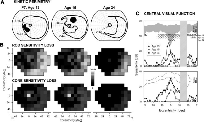
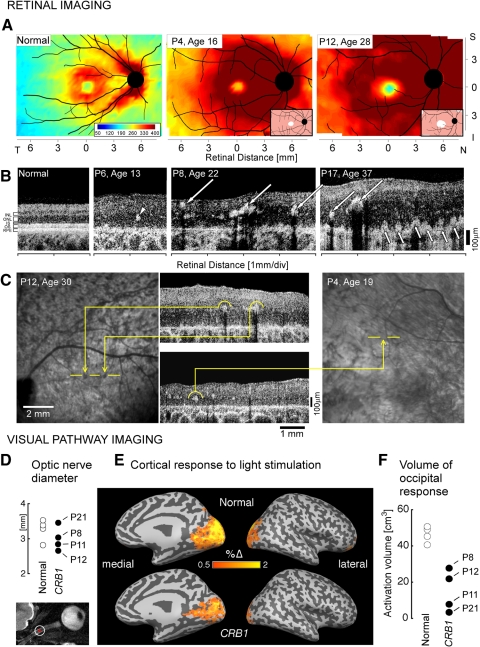
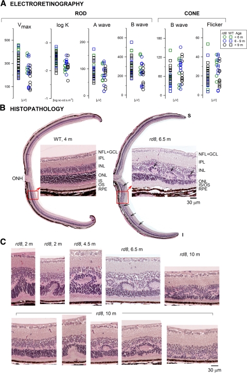
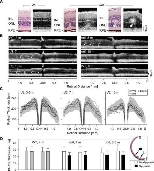
Methods: Twenty-two patients with CRB1 mutations were studied. Function was assessed with perimetry and electroretinography (ERG) and retinal structure with optical coherence tomography (OCT). Cortical structure and function were quantified with magnetic resonance imaging (MRI). Rd8 mice underwent ERG, OCT, and retinal histopathology.

Results: Visual acuities ranged from 20/25 to light perception. Rod ERGs were not detectable; small cone signals were recordable. By perimetry, small central visual islands were separated by midperipheral scotomas from far temporal peripheral islands. The central islands were cone mediated, whereas the peripheral islands retained some rod function. With OCT, there were small foveal islands of thinned outer nuclear layer (ONL) surrounded by thick delaminated retina with intraretinal hyperreflective lesions. MRI showed structurally normal optic nerves and only subtle changes to occipital lobe white and gray matter. Functional MRI indicated that whole-brain responses from patients were of reduced amplitude and spatial extent compared with those of normal controls. Rd8 mice had essentially normal ERGs; OCT and histopathology showed patchy retinal disorganization with pseudorosettes more pronounced in ventral than in dorsal retina. Photoreceptor degeneration was associated with dysplastic regions.

Conclusions: CRB1 mutations lead to early-onset severe loss of vision with thickened, disorganized, nonseeing retina. Impaired peripheral vision can persist in late disease stages. Rd8 mice also have a disorganized retina, but there is sufficient photoreceptor integrity to produce largely normal retinal function. Differences between human and mouse diseases will complicate proof-of-concept studies intended to advance treatment initiatives.


Figures

Figure 1.
Figure 1.
Visual function in representative patients with CRB1-RD. (A) Kinetic perimetry results using two targets (V-4e, I-4e) illustrate preserved central and temporal peripheral islands of vision. (B) Dark-adapted (top) and light-adapted (bottom) static threshold perimetry results displayed as grayscale maps of rod and cone sensitivity loss. The scale has 16 levels of gray, representing 0- to 30-dB losses. N, nasal; T, temporal; I, inferior; S, superior visual field. (C) Dark-adapted, two-color (500 and 650 nm) sensitivity profiles across the horizontal meridian (central 60°) in the patients (symbols connected by lines) compared with normal for rod-mediated sensitivity to the 500-nm stimulus (shaded band) and for cone-mediated sensitivity to the 650 nm stimulus at the cone plateau (dashed lines). The photoreceptor mediation at loci with function, based on the sensitivity difference between the two colors, is given: C, cone-mediated.
Figure 2.
Figure 2.
Longitudinal sequence of visual function in a CRB1-RD patient spanning 11 years. (A) Kinetic perimetry results in P7 at three ages. (B) Dark-adapted (top) and light-adapted (bottom) static threshold perimetry results displayed as grayscale maps of rod and cone sensitivity loss. Scale and labels as in Figure 1. (C) Dark-adapted two-color sensitivity profiles across the central 60° in P7 at the three ages. Top: sensitivity to the 500-nm stimulus compared with normal for rod-mediated sensitivity to this stimulus (shaded band). Bottom: sensitivity to the 650-nm stimulus compared with cone-mediated sensitivity to the same stimulus at the cone plateau (dashed lines). Top graph: photoreceptor mediation at loci with function: M, mixed rod (500 nm) and cone (650 nm) function; C, cone-mediated.
Figure 3.
Figure 3.
Retinal and visual pathway structure in CRB1-RD. (A) Topographical maps of retinal thickness in a normal 21-year-old subject (left) and two patients with CRB1-RD. Traces of major blood vessels and location of the ONH are overlaid on each map. Pseudocolor scales are shown. Insets in the patient maps, bottom right: thickness difference maps showing region that is within normal limits (white, defined as the mean ± 2 SD, n = 5), or thickened (pink, >2 SD), compared to normal. (B) Cross-sectional OCT images along the horizontal meridian in the temporal retina in a normal subject (left, age 24) compared to scans from three patients with CRB1-RD. Arrowhead: intraretinal hyperreflective structures without shadowing. Large arrows: hyperreflective structures with shadowing. Small arrows: hyperreflective lesions apparently extending from RPE vitread into the nuclear layer. (C) En face infrared reflectance images compared with OCT cross sections in two CRB1-RD patients to illustrate the relationship between intraretinal hyperreflective structures, with and without shadowing, and fundus features. In P12, two pigment clumps correspond to intraretinal hyperreflective structures with shadowing. In contrast, intraretinal hyperreflective structures without shadowing in P4 do not correspond to a discrete pigmentary change on the fundus image. (D) High-resolution T2-weighted axial images obtained through the optic nerves. The cross-sectional diameter of the interpial optic nerve was estimated at two positions along each nerve (inset, axial image), and the average diameter was within the range of normal. (E) Cortical (BOLD fMRI) response to light stimulation is shown for normal subjects and CRB1-RD patients on a digitally inflated right hemisphere. Dark gray: sulci; light gray: gyri. The color scale indicates the percentage change of BOLD signal in response to light. Activation in occipital visual cortex is seen for both groups, but is reduced in CRB1-RD. (F) Volume of posterior cortical tissue demonstrating a substantial (2%) response to light stimulation was significantly greater in controls than in CRB1-RD patients.
Figure 4.
Figure 4.
Retinal function and histopathology in the Crb1-mutant rd8 mouse. (A) ERG parameters compared in a cohort of WT and rd8 mice of different ages (key for color coding of ages, right). (B) Dorsal-ventral retinal sections in a 4-month-old WT mouse (left) compared with that of a 6.5-month-old rd8 mouse. To the right of each full retinal section is a higher magnification light microscopic image from a location (square with arrow) inferior to the optic nerve. Arrows: dysplastic regions in the rd8 histopathology. (C) Retinal sections with different abnormalities are ordered (left to right) in a proposed disease sequence leading to the types of dysplastic lesions noted in rd8 animals of increasing ages (top images). Sections are shown from one animal at age 10 months (bottom images) to illustrate that the abnormalities can occur within a retina at a single age.
Figure 5.
Figure 5.
OCT analysis of the retinal abnormalities in rd8 mice. (A) Histologic and OCT sections in WT (left) and rd8 (middle and right) retinas to illustrate the relationship between the different modalities of determining laminar architecture and how abnormalities would appear with noninvasive optical imaging. Left: a 4-month-old WT retina histology section is compared to a 5-month-old WT OCT scan; middle, a 10-month-old rd8 histology section is compared to a 10-month-old OCT image from another rd8 animal; right, 6.5-month-old rd8 histology section is compared to a 7-month-old rd8 OCT scan. (B) Representative OCT scans across 3 mm of retina centered at the ONH, to illustrate detectable abnormalities, such as focal hyperreflective lesions at the level of the OPL (arrowheads) and deeper retinal hyperreflective lesions extending into the ONL (arrows), in the rd8 animals. (C) Dorsal–ventral OCT sections quantified for retinal thickness in three different age groups of rd8 mice. In the 3- to 5-month-old age group, 12 rd8 eyes were analyzed; in the 7-month-old group, 7 eyes; and in the 10-month-old group, 9 rd8 eyes. Shaded bands: the mean ±2 SD of retinal thickness in WT eyes (n = 26, ages 3–8.5 months). Data over this age range were grouped together based on linear regression analyses performed between retinal thickness and age at four selected locations (±0.5 mm and ±1 mm from ONH); slopes of the regression analyses were not significantly different from 0. (D) IS+OS thickness measurements in regions of dysplasia in rd8 histologic sections are compared to adjacent nondysplastic regions (see inset for location of samples) for rd8 mice (two animals each, 4 and 6.5 months old), and in similarly located regions in two WT mice (gray bars). Error bars, ±2 SD.

References

    1. Bazellieres E, Assemat E, Arsanto JP, Le Bivic A, Massey-Harroche D. Crumbs proteins in epithelial morphogenesis. Front Biosci. 2009;14:2149–2169 - PubMed
    1. Heckenlively JR. Preserved para-arteriole retinal pigment epithelium (PPRPE) in retinitis pigmentosa. Br J Ophthalmol. 1982;66:26–30 - PMC - PubMed
    1. den Hollander AI, ten Brink JB, de Kok YJ, et al. Mutations in a human homologue of drosophila crumbs cause retinitis pigmentosa (RP12). Nat Genet. 1999;23:217–221 - PubMed
    1. Lotery AJ, Jacobson SG, Fishman GA, et al. Mutations in the CRB1 gene cause leber congenital amaurosis. Arch Ophthalmol. 2001;119:415–420 - PubMed
    1. Jacobson SG, Cideciyan AV, Aleman TS, et al. Crumbs homolog 1 (CRB1) mutations result in a thick human retina with abnormal lamination. Hum Mol Genet. 2003;12:1073–1078 - PubMed

Publication types

MeSH terms