. 2011 Sep 1;52(10):7059-67.
doi: 10.1167/iovs.11-7497.

Calpain, not caspase, is the causative protease for hypoxic damage in cultured monkey retinal cells

Affiliations

Calpain, not caspase, is the causative protease for hypoxic damage in cultured monkey retinal cells

Emi Nakajima et al. Invest Ophthalmol Vis Sci. .

Abstract

Purpose: Cell death occurring in human retina during AMD, high IOP, and diabetic retinopathy could be caused by activation of calpain or caspase proteolytic enzymes. The purpose of the present study was to determine whether calpains and/or caspase-3 were involved in cell death during retinal hypoxia in a monkey model.

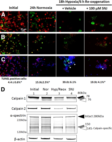
Methods: Dissociated monkey retinal cells were cultured for two weeks and subjected to 24-hour hypoxia/24-hour reoxygenation. TUNEL staining and immunostaining for Müller and photoreceptor markers were used to detect which retinal cell types were damaged.

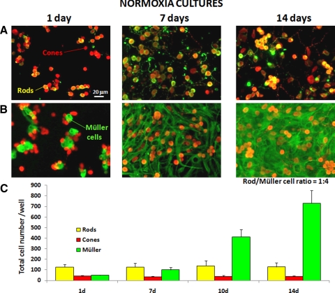
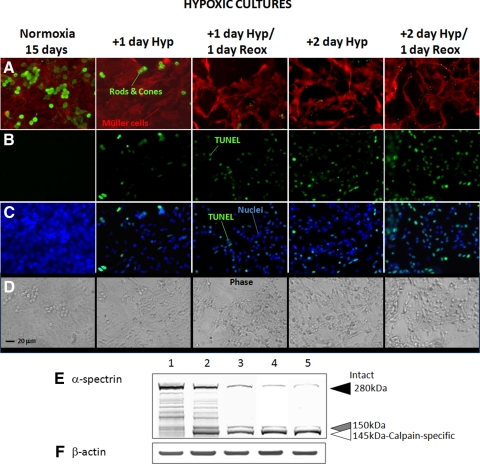
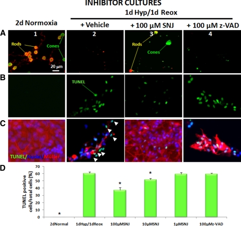
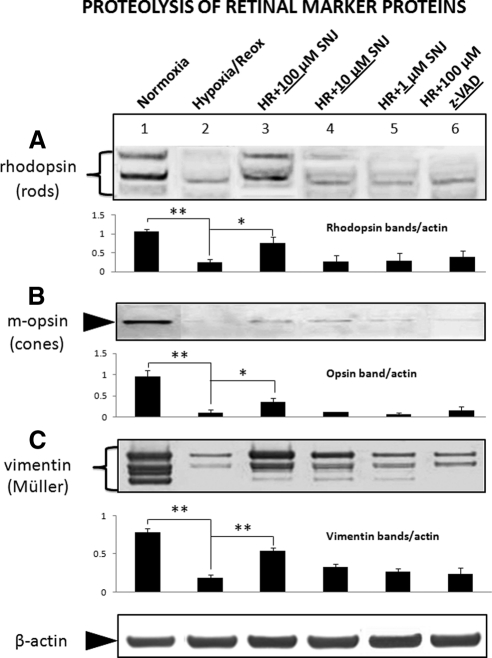
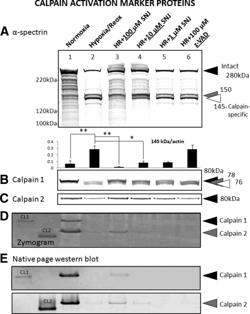
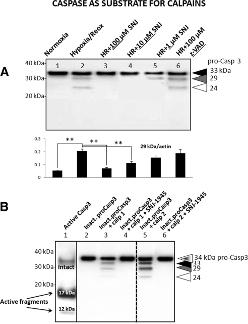
Results: Culturing dissociated monkey retina cells for two weeks resulted in proliferation of Müller cells and maintenance of some rod and cone photoreceptor cells, as identified by vimentin, recoverin, and rhodopsin immunocytochemical staining. Hypoxia/reoxygenation increased the number of cells staining positive for TUNEL. Immunoblotting showed that the calpain-specific 145 kDa α-spectrin breakdown product (SBDP) increased in hypoxic cells, but no caspase-specific 120 kDa α-spectrin breakdown product was detected. TUNEL staining and proteolysis were significantly reduced in the retinal cells treated with 10 and 100 μM calpain inhibitor SNJ-1945. Caspase inhibitor, z-VAD, did not inhibit cell damage from hypoxia/reoxygenation. Intact pro-caspase-3 was in fact cleaved by activated calpain during hypoxia/reoxygenation to pre 29 kDa caspase-3 and 24 kDa inactive fragments. No 17 and 12 kDa fragments, which form the active caspase-3 hetero-dimer, were detected. Calpain-induced cleavage of caspase was inhibited by SNJ-1945.

Conclusions: Calpain, not caspase-3, was involved in hypoxic damage in cultured monkey retinal cells.


Figures

Figure 1.
Figure 1.
Immunofluoresence microscopy of dissociated retinal cells cultured under normoxic conditions for 1, 7, and 14 days showing: (A) rods (yellow, anti-rhodopsin plus anti-recoverin) and cones (red, anti-recoverin); (B) Müller cells (green, anti-vimentin); and (C) counts of cell types expressed as mean ± SEM (n = 3).
Figure 2.
Figure 2.
Immunofluorescence microscopy of dissociated retinal cells cultured under progressively severe hypoxic conditions and quadruple-labeled for cell markers: (A) rods and cones (green, anti-recoverin) and Müller cells (red, anti-vimentin); (B) TUNEL-positive (green) apoptotic cells; (C) image of TUNEL-positive apoptotic cells (green) overlaying image of DAPI-stained nuclei (blue); (D) Phase-contrast micrographs in the stained areas above. (E) Immunoblots for calpain substrate α-spectrin (lane 1) 1 day normal, (lane 2) 1 day hypoxia, (lane 3) 1 day hypoxia/1 day reoxygenation, (lane 4) 2 day hypoxia, (lane 5) 2 day hypoxia/1 day reoxygenation, showing α-spectrin at 280 kDa (filled arrowhead), break down products at 150 (gray arrowhead), and 145 kDa (open arrowhead); (F) Immunoblot for β-actin at 42 kDa as a loading control. Immunofluorescence microscopy and immunoblots are representative of those performed 3 times. Normal groups at days 2 and 3 day were omitted because they showed no changes.
Figure 3.
Figure 3.
Immunofluorescence microscopy of hypoxic retinal cells at 16 days of culture with inhibitors: (lane 3) calpain inhibitor SNJ-1945, or (lane 4) pan-caspase inhibitor z-VAD. (A) Rods (yellow; anti-rhodopsin plus anti-recoverin) and cones (green; anti-recoverin), (B) TUNEL-positive cells (green). (C) Merged images of TUNEL-positive cells (green), DAPI stained nuclei (blue), and Müller cells (red; anti-vimentin) resulting in cyan (arrowheads) labeling of apoptotic cells. (D) Percentages of TUNEL-positive cells. Data are mean ± SEM (n = 3). *P < 0.05 relative to hypoxia/reoxygenation (Dunnett's t-test).
Figure 4.
Figure 4.
Immunoblots for cell-type marker proteins in retinal cells cultured under hypoxic conditions: (lane 2) alone; (lanes 3–5) plus calpain inhibitor; or (lane 6) plus caspase inhibitor. (A) Rhodopsin in rod cells; (B) m-opsin in cone cells; (C) vimentin in Müller cells. Bar graphs indicate band intensity normalized to β-actin loading control, expressed as mean ± SEM (n = 3 to 6). *P < 0.05, **P < 0.01, all relative to hypoxia/reoxygenation (lane 2), using Dunnett's t-test.
Figure 5.
Figure 5.
Immunoblots of marker proteins indicating calpain activation in hypoxic retinal cells cultured: (lane 2) alone; (lanes 3–5) plus calpain inhibitor; or (lane 6) plus caspase inhibitor. (A) Intact α-spectrin at 280 kDa (filled arrowhead) and breakdown products at 150 kDa (gray arrowhead) and the calpain-specific fragment at 145 kDa (open arrowhead). Below: density of α-spectrin 145 kDa band normalized to the β-actin loading control, expressed as mean ± SEM (n = 3 to 6) *P < 0.05, **P < 0.01, all relative to hypoxia/reoxygenation (lane 2), using Dunnett's t-test. (B) Intact calpain 1 catalytic subunit at 80 kDa (black arrowhead) and fragments at 78 kDa (gray arrowhead) and 76 kDa (open arrowhead) observed after autolytic activation. (C) Intact calpain 2 catalytic subunit at 80 kDa (black arrowhead). Autolyzed, active form of calpain 2 shows the same migration as intact 80 kDa calpain 2 on SDS-PAGE., (D) Casein zymogram (image inverted) showing remaining active calpains in the cultured retinal samples (20 μg per lane). Black arrowhead indicates active calpain 1, and gray arrowhead indicates active calpain 2. The left two lanes are purified human erythrocyte calpain 1 (CL1) and porcine kidney calpain 2 (CL2) used as standards and positive controls. (E) Immunoblots of calpain 1 and calpain 2 from native PAGE gel used to confirm migration positions of active calpains 1 and 2 in the cultured retinal samples (20 μg per lane). The left two lanes are purified human erythrocyte calpain 1 (CL1) and porcine kidney calpain 2 (CL2) used as standards and positive controls.
Figure 6.
Figure 6.
Immunofluorescence microscopy of monkey retina cells in suspension culture after 24 hours of hypoxia. (A) Vimentin (green) and rods and cones (red; anti-recoverin), (B) rods (yellow; anti-rhodopsin and anti-recoverin) and cones (green; anti-recoverin), (C) merged images of TUNEL-positive cells (green), DAPI-stained nuclei (blue), and rods and cones (red; anti-recoverin) resulting in cyan (arrowheads) labeling of apoptotic cells (same areas as in B). Percentages of TUNEL-positive cells are shown below the images. Data are mean ± SEM (n = 3). *P < 0.05 relative to hypoxia/reoxygenation (Dunnett's t-test). (D) Immunoblots for marker proteins for calpain activation: calpain 1, calpain 2, α-spectrin; β-actin as loading control.
Figure 7.
Figure 7.
(A) Immunoblot for endogenous pro-caspase-3 in hypoxic retinal cells treated without (lane 2) and with enzyme inhibitors (lanes 3–6). Intact pro-caspase-3 is indicated at 33 kDa (filled arrowhead) along with calpain-dependent breakdown products at 29 (gray arrowhead) and 24 kDa (open arrowhead). Below: density of caspase-3 bands at 29 and 24 kDa normalized to β-actin loading control, expressed as mean ± SEM (n = 3 to 9), **P < 0.01 all relative to the hypoxia/reoxygenation group (lane 2) using Dunnett's t-test. (B) Immunoblots for pro-capsase 3 after in vitro incubation of: (lane 1) activated recombinant caspase-3 showing loss of intact pro-caspase-3 at 34 kDa, production of known active caspase-3 fragments at 17 and 12 kDa (arrows), and absence of inactive 29 and 24 kDa fragments. Other lanes show: (lane 2) incubation of inactive recombinant pro-caspase-3 alone; (lane 3) plus purified active calpain 1; (lane 4) plus active calpain 1 and 100 μM calpain inhibitor; (lane 5) plus purified active calpain 2; or (lane 6) plus active calpain 2 and 100 μM calpain inhibitor.

Similar articles

Cited by

References

    1. Azuma M, Shearer TR. The role of calcium-activated protease calpain in experimental retinal pathology. Surv Ophthalmol. 2008;53:150–163 - PMC - PubMed
    1. Kawasaki A, Otori Y, Barnstable CJ. Muller cell protection of rat retinal ganglion cells from glutamate and nitric oxide neurotoxicity. Invest Ophthalmol Vis Sci. 2000;41:3444–3450 - PubMed
    1. Bringmann A, Pannicke T, Grosche J, et al. Muller cells in the healthy and diseased retina. Prog Retin Eye Res. 2006;25:397–424 - PubMed
    1. Das AV, Mallya KB, Zhao X, et al. Neural stem cell properties of Müller glia in the mammalian retina: regulation by Notch and Wnt signaling. Dev Biol. 2006;299:283–302 - PubMed
    1. Lawrence JM, Singhal S, Bhatia B, et al. MIO-M1 cells and similar Muller glial cell lines derived from adult human retina exhibit neural stem cell characteristics. Stem Cells. 2007:25;2033–2043 - PubMed

Publication types

MeSH terms